Application
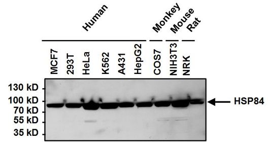
WB analysis of indicated cell lysates (50ug per lane) HSP84 antibody at a dilution of 1:1000.Application
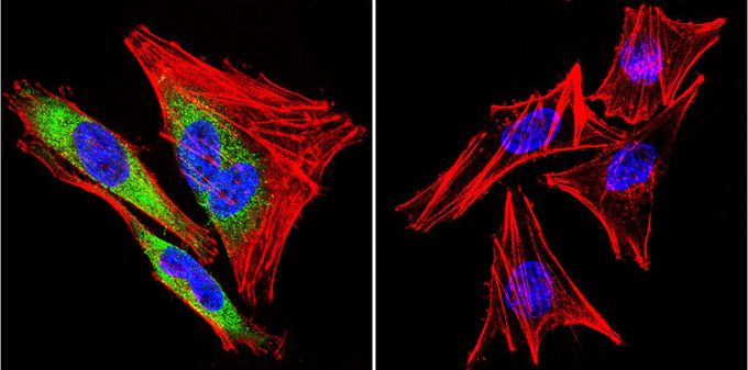
ICC/IF analysis of A2058 cells with (left) or without (right) HSP84 antibody at a dilution of 1:100 (green). F-Actin staining with Phalloidin (red) and nuclei with DAPI (blue) is shown. Images were taken at 60X magnification.Application
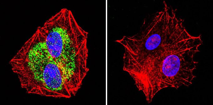
ICC/IF analysis of HepG2 cells with (left) or without (right) using HSP84 antibody at a dilution of 1:100 (green). F-Actin staining with Phalloidin (red) and nuclei with DAPI (blue) is shown. Images were taken at 60X magnification.Application
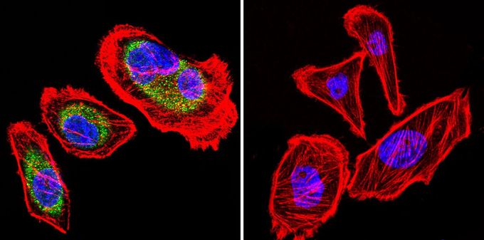
ICC/IF analysis of U251 cells with (green) or without (left) HSP84 antibody at a dilution of 1:100 (green). F-Actin staining with Phalloidin (red) and nuclei with DAPI (blue) is shown. Images were taken at 60X magnification.Application
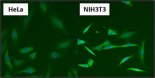
ICC/IF analysis of HeLa and NIH3T3 cells using HSP84 antibody at a dilution of 1:50 (green) and Hoechst 33342 (blue). Images were taken at 20X magnification.Application
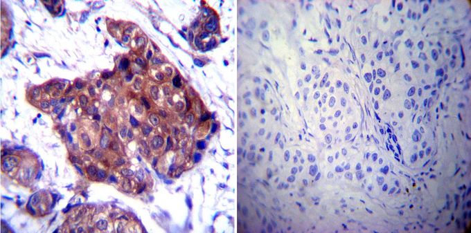
IHC-P analysis of human breast carcinoma with (left) or without (right) HSP84 antibody at a dilution of 1:100. To expose target proteins, heat induced antigen retrieval was performed using 10mM sodium citrate (pH6.0) buffer, microwaved for 8-15 minutes.Application
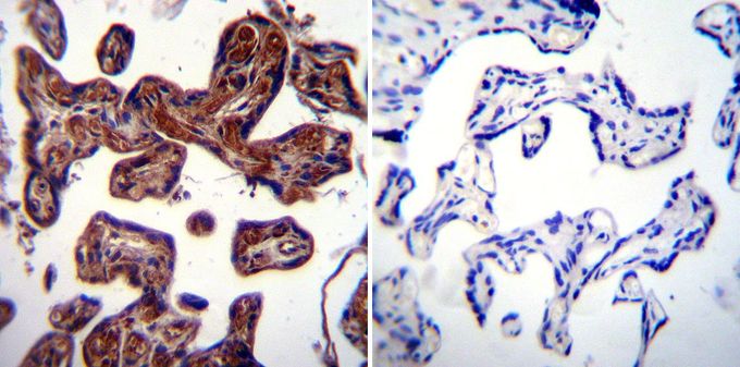
IHC-P analysis of human placenta tissue with (left) or without (right) HSP84 antibody at a dilution of 1:20. To expose target proteins, heat induced antigen retrieval was performed using 10mM sodium citrate (pH6.0) buffer, microwaved for 8-15 minutes.Application
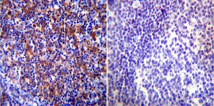
IHC-P analysis of human tonsil tissue with (left) or without (right) HSP84 antibody at a dilution of 1:100. To expose target proteins, heat induced antigen retrieval was performed using 10mM sodium citrate (pH6.0) buffer, microwaved for 8-15 minutes.Application
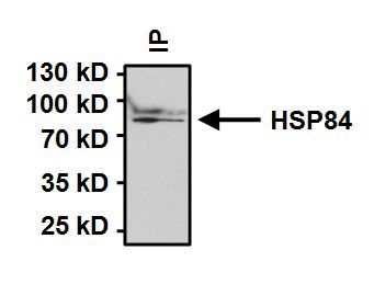
IP analysis was performed on 500 ug of HeLa extract using 2 ug of HSP84 antibody. The precipitated protein was detected by the same antibody at a dilution of 1:1000.| Product Name | Heat Shock Protein 84 antibody |
|---|---|
| Antibody Type | Primary Antibodies |
| Product description | Rabbit Polyclonal antibody to Heat Shock Protein 84Heat shock proteins (HSP) are expressed in response to various biological stresses, including heat. HSP90 is a 90 kDa protein that is induced under stress conditions, but is also one of the most abundant cellular proteins found under non-stress conditions. HSP90 has been found to be associated with a number of other intracellular proteins, including steroid receptors, actin, tubulin, aryl hydrocarbon (Ah) receptor, and some kinases. Studies have shown that murine HSP90 exists as two forms, HSP84 and HSP86, coded by related but separate genes, with 86% amino acid sequence conservation. These forms are analogous to the two forms of human HSP90, HSP89 alpha and HSP89 beta. In an unstressed mouse fibroblast, the basal level of HSP84 is found to be double that of HSP86. However, after heat shock, HSP86 shows a greater increase. Studies also suggest that upon cellular differentiation, the level of HSP86, but not HSP84, decreases. HSP84 and HSP86, which may be subject to estrogenic regulation, have been found as components of the non-DNA binding form of mouse glucocorticoid receptor, but dissociated from the transformed DNA-binding form. |
| Immunogen | Synthetic peptide corresponding to residues P(2) E E V H H G E E E V E(13) of mouse HSP84. |
| Clonality | Polyclonal |
|---|---|
| Host Species | Rabbit |
| Tested Applications | ICC/IFIHCIHC-FIHC-PIPWB |
| ICC/IF: 10 - 20 µg/ml: |
|
| Species Reactivity | HumanMouseRatPrimate |
| Concentration | 1mg/ml (Please refer to the vial label for the specific concentration) |
| Purification | Affinity purified |
| Gene Synonyms | Hsp90ab1 90kDa AL022974 C81438 Hsp84 Hsp84-1 Hsp90 Hspcb MGC115780 heat shock protein 90 alpha (cytosolic) class B member 1 |
|---|---|
| Target | Heat Shock Protein 84 |
Application
WB analysis of indicated cell lysates (50ug per lane) HSP84 antibody at a dilution of 1:1000.
Application
ICC/IF analysis of A2058 cells with (left) or without (right) HSP84 antibody at a dilution of 1:100 (green). F-Actin staining with Phalloidin (red) and nuclei with DAPI (blue) is shown. Images were taken at 60X magnification.
Application
ICC/IF analysis of HepG2 cells with (left) or without (right) using HSP84 antibody at a dilution of 1:100 (green). F-Actin staining with Phalloidin (red) and nuclei with DAPI (blue) is shown. Images were taken at 60X magnification.
Application
ICC/IF analysis of U251 cells with (green) or without (left) HSP84 antibody at a dilution of 1:100 (green). F-Actin staining with Phalloidin (red) and nuclei with DAPI (blue) is shown. Images were taken at 60X magnification.
Application
ICC/IF analysis of HeLa and NIH3T3 cells using HSP84 antibody at a dilution of 1:50 (green) and Hoechst 33342 (blue). Images were taken at 20X magnification.
Application
IHC-P analysis of human breast carcinoma with (left) or without (right) HSP84 antibody at a dilution of 1:100. To expose target proteins, heat induced antigen retrieval was performed using 10mM sodium citrate (pH6.0) buffer, microwaved for 8-15 minutes.
Application
IHC-P analysis of human placenta tissue with (left) or without (right) HSP84 antibody at a dilution of 1:20. To expose target proteins, heat induced antigen retrieval was performed using 10mM sodium citrate (pH6.0) buffer, microwaved for 8-15 minutes.
Application
IHC-P analysis of human tonsil tissue with (left) or without (right) HSP84 antibody at a dilution of 1:100. To expose target proteins, heat induced antigen retrieval was performed using 10mM sodium citrate (pH6.0) buffer, microwaved for 8-15 minutes.
Application
IP analysis was performed on 500 ug of HeLa extract using 2 ug of HSP84 antibody. The precipitated protein was detected by the same antibody at a dilution of 1:1000.| Application Notes | ICC/IF: 10 - 20 µg/ml: |
|---|
| Form | Liquid |
|---|---|
| Storage Instructions | Keep as concentrated solution. Aliquot and store at -20ºC or below. Avoid multiple freeze-thaw cycles. |
| Storage Buffer | PBS containing 1 mg/ml BSA and 0.05% sodium azide. |


