Application
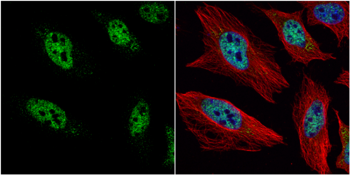
HES1 antibody [N1], N-term detects HES1 protein at nucleus by immunofluorescent analysis. Sample: HeLa cells were fixed in 4% paraformaldehyde at RT for 15 min. Green: HES1 protein stained by HES1 antibody [N1], N-term diluted at 1:2000. Red: alpha Tubulin, a cytoskeleton marker, stained by alpha Tubulin antibody [GT114] diluted at 1:1000. Blue: Hoechst 33342 staining.Application
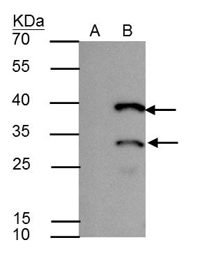
HES1 antibody immunoprecipitates HES1 protein in IP experiments. IP Sample: A431 whole cell lysate/extract A : Control with 2.5 μg of pre-immune rabbit IgG B : Immunoprecipitation of HES1 by 2.5 μg of HES1 antibody 12% SDS-PAGE The immunoprecipitated HES1 protein was detected by HES1 antibody diluted at 1 : 1000. EasyBlot anti-rabbit IgG (HRP) was used as a secondary reagent.Application
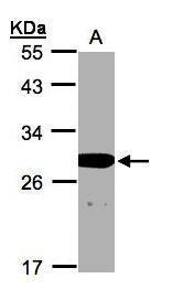
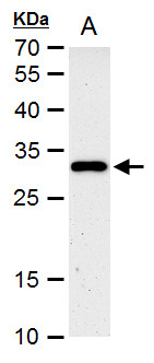
Sample(30 µg whole cell lysate) A:A431 12% SDS PAGE diluted at 1:1000 The HRP-conjugated anti-rabbit IgG antibody was used to detect the primary antibody.Application
HES1 antibody [N1], N-term detects HES1 protein by western blot analysis. A.50 µg mouse kidney extract 12% SDS-PAGE HES1 antibody [N1], N-term dilution: 1:1000 The HRP-conjugated anti-rabbit IgG antibody was used to detect the primary antibody.Application
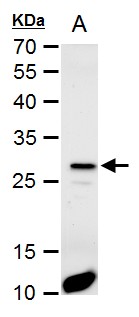
HES1 antibody [N1], N-term detects HES1 protein by western blot analysis. A. 30 µg PC-12 whole cell extract 12% SDS-PAGE HES1 antibody [N1], N-term dilution: 1:1000 The HRP-conjugated anti-rabbit IgG antibody was used to detect the primary antibody.| Product Name | HES1 antibody , N-term |
|---|---|
| Antibody Type | Primary Antibodies |
| Product description | Rabbit polyclonal antibody to HES1 (hairy and enhancer of split 1, (Drosophila))This protein belongs to the basic helix-loop-helix family of transcription factors. It is a transcriptional repressor of genes that require a bHLH protein for their transcription. The protein has a particular type of basic domain that contains a helix interrupting protein that binds to the N-box rather than the canonical E-box. [provided by RefSeq] |
| Immunogen | Carrier-protein conjugated synthetic peptide encompassing a sequence within the N-terminus region of human HES1. The exact sequence is proprietary. |
| Clonality | Polyclonal |
|---|---|
| Isotype | IgG |
| Host Species | Rabbit |
| Tested Applications | ICC/IFIPWB |
| *Optimal dilutions/concentrations should be determined by the researcher.
: |
|
| Species Reactivity | HumanMouseRat |
| Predicted Reactivity | Pig, BovinePig (100%), Bovine (100%) |
| Concentration | 1mg/ml (Please refer to the vial label for the specific concentration) |
| Purification | Affinity purified |
| Gene Synonyms | FLJ20408 antibody HES-1 antibody HHL antibody HRY antibody bHLHb39 antibody HES1 antibody hairy-like protein antibody hairy homolog antibody class B basic helix-loop-helix protein 39 antibody transcription factor HES-1 antibody "hairy and enhance |
|---|---|
| Target | HES1 |
| Molecular Weight(MW) | 30 kDa (note) |
| Cellular Localization | Nucleus |
Application
HES1 antibody [N1], N-term detects HES1 protein at nucleus by immunofluorescent analysis. Sample: HeLa cells were fixed in 4% paraformaldehyde at RT for 15 min. Green: HES1 protein stained by HES1 antibody [N1], N-term diluted at 1:2000. Red: alpha Tubulin, a cytoskeleton marker, stained by alpha Tubulin antibody [GT114] diluted at 1:1000. Blue: Hoechst 33342 staining.
Application
HES1 antibody immunoprecipitates HES1 protein in IP experiments. IP Sample: A431 whole cell lysate/extract A : Control with 2.5 μg of pre-immune rabbit IgG B : Immunoprecipitation of HES1 by 2.5 μg of HES1 antibody 12% SDS-PAGE The immunoprecipitated HES1 protein was detected by HES1 antibody diluted at 1 : 1000. EasyBlot anti-rabbit IgG (HRP) was used as a secondary reagent.
Application
Sample(30 µg whole cell lysate) A:A431 12% SDS PAGE diluted at 1:1000 The HRP-conjugated anti-rabbit IgG antibody was used to detect the primary antibody.
Application
HES1 antibody [N1], N-term detects HES1 protein by western blot analysis. A.50 µg mouse kidney extract 12% SDS-PAGE HES1 antibody [N1], N-term dilution: 1:1000 The HRP-conjugated anti-rabbit IgG antibody was used to detect the primary antibody.
Application
HES1 antibody [N1], N-term detects HES1 protein by western blot analysis. A. 30 µg PC-12 whole cell extract 12% SDS-PAGE HES1 antibody [N1], N-term dilution: 1:1000 The HRP-conjugated anti-rabbit IgG antibody was used to detect the primary antibody.| Positive Control | 293T , A431 , H1299 , HeLaS3 , HepG2 , Molt-4 , Raji , mouse kidney , PC-12 |
|---|---|
| Application Notes | *Optimal dilutions/concentrations should be determined by the researcher.
: |
| Form | Liquid |
|---|---|
| Storage Instructions | Keep as concentrated solution. Aliquot and store at -20ºC or below. Avoid multiple freeze-thaw cycles. |
| Storage Buffer | 0.1M Tris, 0.1M Glycine, 10% Glycerol (pH7). 0.01% Thimerosal was added as a preservative. |


