Application
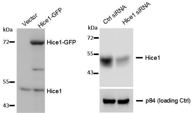
WB to detect cellular Hice1 and Hice1-GFP expressed in human osteosarcoma U2OS cells (left image), and Hice1 upon siRNA treatment (right image), using at 1:1000 dilution. Nuclear matrix protein p84 is a loading control, blotted with p84 antibody (clone 5E10) at 1:5000 dilution. The HRP-conjugated anti-rabbit IgG antibody was used to detect the primary antibody.Application
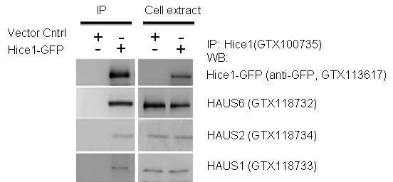
IP-WB assay to show that Hice1 co-immunoprecipitated with other Augmin components HAUS6 , HAUS2 and HAUS1 in U2OS cells.Application
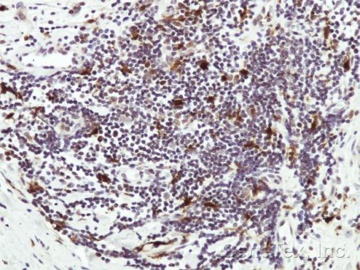
Immunohistochemical analysis of paraffin-embedded Human lymph tissue, using HICE1 antibody at 1:50 dilution.Application
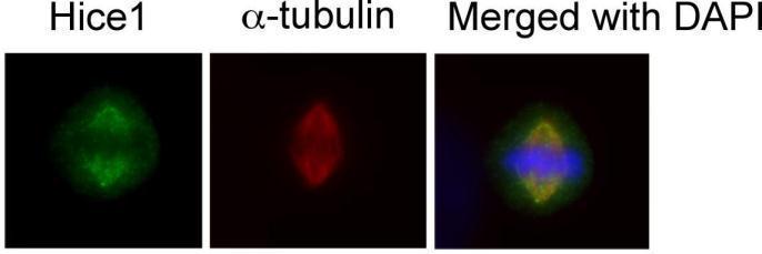
Immunofluorescence analysis of human osteosarcoma cell line U2OS, using HICE1 antibody at 1:50-1:200 dilution.Application
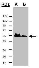
Sample A: His-Hice1 (2x) B: His-Hice1 (1x) 7.5% SDS PAGE diluted at 1:500 The HRP-conjugated anti-rabbit IgG antibody was used to detect the primary antibody.| Product Name | HICE1 antibody |
|---|---|
| Antibody Type | Primary Antibodies |
| Product description | Rabbit polyclonal antibody to HICE1 (HAUS augmin-like complex, subunit 8)HAUS8 is 1 of 8 subunits of the 390-kD human augmin complex, or HAUS complex. The augmin complex was first identified in Drosophila, and its name comes from the Latin verb 'augmentare,' meaning 'to increase.' The augmin complex is a microtubule-binding complex involved in microtubule generation within the mitotic spindle and is vital to mitotic spindle assembly (Goshima et al., 2008 [PubMed 18443220]; Uehara et al., 2009 [PubMed 19369198]).[supplied by OMIM] |
| Clonality | Polyclonal |
|---|---|
| Isotype | IgG |
| Host Species | Rabbit |
| Tested Applications | ICC/IFIHC-PIPWB |
| *Optimal dilutions/concentrations should be determined by the researcher.
: |
|
| Species Reactivity | Human |
| Concentration | 1mg/ml |
| Purification | Affinity purified |
| Gene Synonyms | DGT4 antibody HICE1 antibody MGC20533 antibody NY-SAR-48 antibody HAUS8 antibody "HEC1/NDC80 interacting centrosome associated 1 antibody" Hec1-interacting and centrosome-associated 1 antibody sarcoma antigen NY-SAR-48 antibody HEC1/NDC80-interact |
|---|---|
| Target | HICE1 |
| Molecular Weight(MW) | 45 kDa (note) |
| Cellular Localization | Cytoplasm , Centrosome , Spindle |
Application
WB to detect cellular Hice1 and Hice1-GFP expressed in human osteosarcoma U2OS cells (left image), and Hice1 upon siRNA treatment (right image), using at 1:1000 dilution. Nuclear matrix protein p84 is a loading control, blotted with p84 antibody (clone 5E10) at 1:5000 dilution. The HRP-conjugated anti-rabbit IgG antibody was used to detect the primary antibody.
Application
IP-WB assay to show that Hice1 co-immunoprecipitated with other Augmin components HAUS6 , HAUS2 and HAUS1 in U2OS cells.
Application
Immunohistochemical analysis of paraffin-embedded Human lymph tissue, using HICE1 antibody at 1:50 dilution.
Application
Immunofluorescence analysis of human osteosarcoma cell line U2OS, using HICE1 antibody at 1:50-1:200 dilution.
Application
Sample A: His-Hice1 (2x) B: His-Hice1 (1x) 7.5% SDS PAGE diluted at 1:500 The HRP-conjugated anti-rabbit IgG antibody was used to detect the primary antibody.| Positive Control | K562 , HeLa |
|---|---|
| Application Notes | *Optimal dilutions/concentrations should be determined by the researcher.
: |
| Form | Liquid |
|---|---|
| Storage Instructions | Keep as concentrated solution. Aliquot and store at -20ºC or below. Avoid multiple freeze-thaw cycles. |
| Storage Buffer | 0.1M Tris, 0.1M Glycine, 10% Glycerol (pH7). 0.01% Thimerosal was added as a preservative. |


