Application
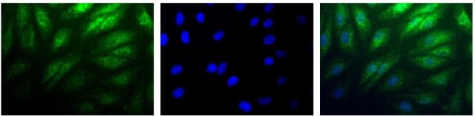
Immunocytochemistry/Immunofluorescence: HIF-2 alpha Antibody - Detection of HIF-2 Alpha (Green) in RCC4 cells using . Nuclei (Blue) are counterstained with Hoechst 33258.Application
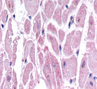
Immunohistochemistry: HIF-2 alpha Antibody - Hif-2 alpha immunoreactivity in human cardiac myocytesApplication
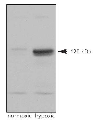
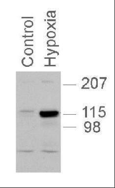
Western Blot: HIF-2 alpha Antibody - Analysis on normoxic and hypoxic nuclear rat cell lysates.Application
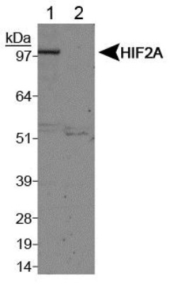
Western Blot: HIF-2 alpha Antibody - Analysis of HIF-2 alpha on Lane 1, Cobalt chloride treated COS7 nuclear extracts and Lane 2, Untreated COS7 nuclear extracts usingApplication
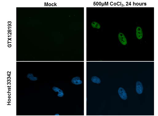
HIF2 alpha antibody detects HIF2 alpha protein at nucleus by immunofluorescent analysis. Sample: HeLa cells were fixed in 4% paraformaldehyde at RT for 15 min. Green: HIF2 alpha protein stained by HIF2 alpha antibody diluted at 1:200. Blue: Hoechst 33342 staining.Application
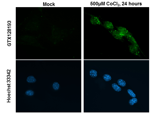
HIF2 alpha antibody detects HIF2 alpha protein at nucleus by immunofluorescent analysis. Sample: NIH/3T3 cells were fixed in 4% paraformaldehyde at RT for 15 min. Green: HIF2 alpha protein stained by HIF2 alpha antibody diluted at 1:200. Blue: Hoechst 33342 staining.Application
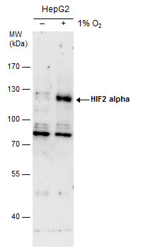
HIF2 alpha antibody detects HIF2 alpha protein by western blot analysis. Un-treated (-) and treated (+, 0.01 O2 treatment for 24h) HepG2 whole cell extracts (30 µg) were separated by 7.5% SDS-PAGE, and the membrane was blotted with HIF2 alpha antibody diluted by 1:500. The HRP-conjugated anti-rabbit IgG antibody was used to detect the primary antibody.Application
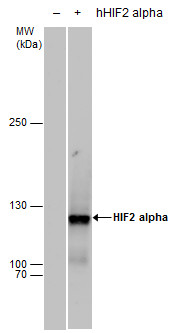
HIF2 alpha antibody detects HIF2 alpha protein by western blot analysis. Non-transfected (-) and HIF2A-transfected (+, including Myc-DDK-tag) 293T whole cell extracts (30 µg) were separated by 5% SDS-PAGE, and the membrane was blotted with HIF2 alpha antibody diluted by 1:1000. The HRP-conjugated anti-rabbit IgG antibody was used to detect the primary antibody.Application
utilized in western blot with PC12 nuclear extracts, 1:1,000, 5 second exposure.| Product Name | HIF2 alpha antibody |
|---|---|
| Antibody Type | Primary Antibodies |
| Product description | Rabbit polyclonal to HIF - 2 alpha (EPAS1)Hypoxia contributes significantly to the pathophysiology of major categories of human disease, including myocardial and cerebral ischemia, cancer, pulmonary hypertension, congenital heart disease and chronic obstructive pulmonary disease. HIF-2 alpha is predominantly expressed in highly vascularized tissues of adult humans and endothelial cells of the embryonic and adult mouse, whereas HIF-1alpha functions primarily in extravascular tissues. HIF-2 alpha is also a potent activator of the tie-2 gene, which is known to be selectively expressed in endothelial cells. (1) |
| Immunogen | A peptide derived from the mouse/human HIF-2 alpha. |
| Clonality | Polyclonal |
|---|---|
| Isotype | IgG |
| Host Species | Rabbit |
| Tested Applications | ICC/IFIHCWB |
| *Optimal dilutions/concentrations should be determined by the researcher.
: |
|
| Species Reactivity | HumanMouseRatFish |
| Concentration | 1mg/ml (Please refer to the vial label for the specific concentration) |
| Purification | Affinity purified |
| Gene Synonyms | Epas1 13819 HIF2 alpha HIF2alpha HIF-2alpha HLF MOP2 HIF 2alpha |
|---|---|
| Target | HIF2 alpha |
| Tissue Specificity | Specific for HIF-2 alpha/EPAS. Does not cross react with HIF-1 alpha. |
Application
Immunocytochemistry/Immunofluorescence: HIF-2 alpha Antibody - Detection of HIF-2 Alpha (Green) in RCC4 cells using . Nuclei (Blue) are counterstained with Hoechst 33258.
Application
Immunohistochemistry: HIF-2 alpha Antibody - Hif-2 alpha immunoreactivity in human cardiac myocytes
Application
Western Blot: HIF-2 alpha Antibody - Analysis on normoxic and hypoxic nuclear rat cell lysates.
Application
Western Blot: HIF-2 alpha Antibody - Analysis of HIF-2 alpha on Lane 1, Cobalt chloride treated COS7 nuclear extracts and Lane 2, Untreated COS7 nuclear extracts using
Application
HIF2 alpha antibody detects HIF2 alpha protein at nucleus by immunofluorescent analysis. Sample: HeLa cells were fixed in 4% paraformaldehyde at RT for 15 min. Green: HIF2 alpha protein stained by HIF2 alpha antibody diluted at 1:200. Blue: Hoechst 33342 staining.
Application
HIF2 alpha antibody detects HIF2 alpha protein at nucleus by immunofluorescent analysis. Sample: NIH/3T3 cells were fixed in 4% paraformaldehyde at RT for 15 min. Green: HIF2 alpha protein stained by HIF2 alpha antibody diluted at 1:200. Blue: Hoechst 33342 staining.
Application
HIF2 alpha antibody detects HIF2 alpha protein by western blot analysis. Un-treated (-) and treated (+, 0.01 O2 treatment for 24h) HepG2 whole cell extracts (30 µg) were separated by 7.5% SDS-PAGE, and the membrane was blotted with HIF2 alpha antibody diluted by 1:500. The HRP-conjugated anti-rabbit IgG antibody was used to detect the primary antibody.
Application
HIF2 alpha antibody detects HIF2 alpha protein by western blot analysis. Non-transfected (-) and HIF2A-transfected (+, including Myc-DDK-tag) 293T whole cell extracts (30 µg) were separated by 5% SDS-PAGE, and the membrane was blotted with HIF2 alpha antibody diluted by 1:1000. The HRP-conjugated anti-rabbit IgG antibody was used to detect the primary antibody.
Application
utilized in western blot with PC12 nuclear extracts, 1:1,000, 5 second exposure.| Positive Control | Hypoxic lysates , HepG2 (1% O2 treatment for 24hr and using Urea buffer) , Myc-DDK human HIF2A-transfected 293T |
|---|---|
| Application Notes | *Optimal dilutions/concentrations should be determined by the researcher.
: |
| Form | Liquid |
|---|---|
| Storage Instructions | Keep as concentrated solution. Aliquot and store at -20ºC or below. Avoid multiple freeze-thaw cycles. |
| Storage Buffer | 1XPBS, 20% Glycerol (pH7). 0.01% Thimerosal was added as a preservative. |


