Application
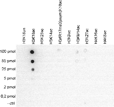
Dot blot analysis of peptides containing histone modifications and the unmodified H3K18 using H3K18ac antibody at a dilution of 1:5,000. One hundred to 0.2 pmol of the respective peptides were spotted on a membrane for analysis.Application
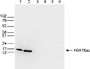
WB analysis of whole cell (25 μg, lane 1) and histone extracts (15 μg, lane 2) from HeLa cells, and 1 μg of recombinant histone H2A, H2B, H3 and H4 (lane 3, 4, 5 and 6, respectively) using H3K18ac antibody at a dilution of 1:500 in TBS-Tween containing 5% skimmed milk.Application
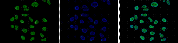
ICC/IF analysis of HeLa cells using H3K18ac antibody (green) and DAPI (blue). Cells were fixed with 4% formaldehyde for 10’ and blocked with PBS/TX-100 containing 5% normal goat serum and 1% BSA. The cells were immunofluorescently labeled with H3K18ac antibody at a dilution of 1:200.Application
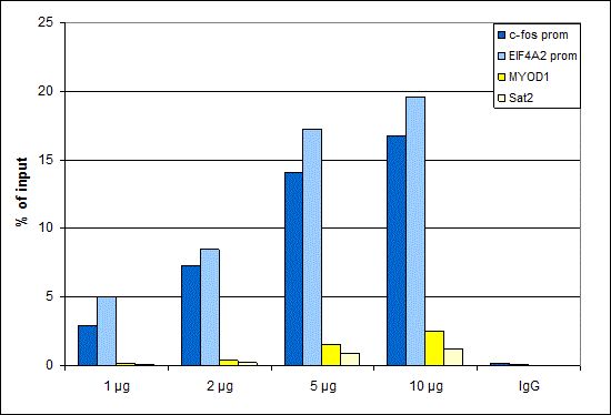
ChIP was performed with extracts from TSA-treated HeLa cells and either control IgG (2 μg) or Histone H3K18ac (acetyl Lys18) antibody (1, 2, 5 and 10 μg per ChIP experiment). The precipitated DNA was detected by qPCR with primers targeting to positive controls c-fos and EIF4A2 promoters, and to negative controls MYOD1 gene and the Sat2 satellite repeat.Application
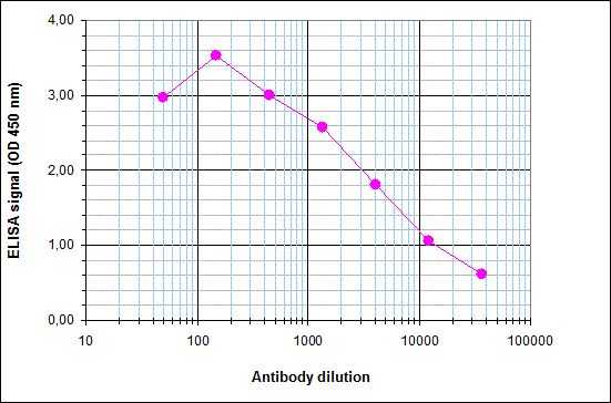
ELISA was performed using a serial dilution of Histone H3K18ac (acetyl Lys18) antibody in antigen coated wells. By plotting the absorbance against the antibody dilution, the titer of the antibody was estimated to be 1:4,300| Product Name | Histone H3K18ac (acetyl Lys18) antibody - ChIP grade |
|---|---|
| Antibody Type | Primary Antibodies |
| Product description | Rabbit polyclonal antibody to H3 (acetyl Lys18)Histones are basic nuclear proteins that are responsible for the nucleosome structure of the chromosomal fiber in eukaryotes. This structure consists of approximately 146 bp of DNA wrapped around a nucleosome, an octamer composed of pairs of each of the four core histones (H2A, H2B, H3, and H4). The chromatin fiber is further compacted through the interaction of a linker histone, H1, with the DNA between the nucleosomes to form higher order chromatin structures. This gene is intronless and encodes a member of the histone H3 family. Transcripts from this gene lack polyA tails; instead, they contain a palindromic termination element. This gene is found in the large histone gene cluster on chromosome 6p22-p21.3. [provided by RefSeq, Jul 2008] |
| Immunogen | Polyclonal antibody raised in rabbit against the region of histone H3 containing the acetylated lysine 18 (H3K18ac), using a KLH-conjugated synthetic peptide. |
| Modification | acetyl Lys18 |
| Clonality | Polyclonal |
|---|---|
| Host Species | Rabbit |
| Tested Applications | DotELISAICC/IFWBChIP |
| ChIP/ChIP-seq (1 µg/IP), ELISA (1:100), Dot blotting (1:5,000), Western blotting (1:500), Immunofluorescence (1:200): |
|
| Species Reactivity | HumanMouse |
| Concentration | 1mg/ml |
| Purification | Affinity purified |
| Gene Synonyms | histone cluster 1 H3a H3/A HIST1H3A H3FA |
|---|---|
| Target | H3 (acetyl Lys18) |
Application
Dot blot analysis of peptides containing histone modifications and the unmodified H3K18 using H3K18ac antibody at a dilution of 1:5,000. One hundred to 0.2 pmol of the respective peptides were spotted on a membrane for analysis.
Application
WB analysis of whole cell (25 μg, lane 1) and histone extracts (15 μg, lane 2) from HeLa cells, and 1 μg of recombinant histone H2A, H2B, H3 and H4 (lane 3, 4, 5 and 6, respectively) using H3K18ac antibody at a dilution of 1:500 in TBS-Tween containing 5% skimmed milk.
Application
ICC/IF analysis of HeLa cells using H3K18ac antibody (green) and DAPI (blue). Cells were fixed with 4% formaldehyde for 10’ and blocked with PBS/TX-100 containing 5% normal goat serum and 1% BSA. The cells were immunofluorescently labeled with H3K18ac antibody at a dilution of 1:200.
Application
ChIP was performed with extracts from TSA-treated HeLa cells and either control IgG (2 μg) or Histone H3K18ac (acetyl Lys18) antibody (1, 2, 5 and 10 μg per ChIP experiment). The precipitated DNA was detected by qPCR with primers targeting to positive controls c-fos and EIF4A2 promoters, and to negative controls MYOD1 gene and the Sat2 satellite repeat.
Application
ELISA was performed using a serial dilution of Histone H3K18ac (acetyl Lys18) antibody in antigen coated wells. By plotting the absorbance against the antibody dilution, the titer of the antibody was estimated to be 1:4,300| Application Notes | ChIP/ChIP-seq (1 µg/IP), ELISA (1:100), Dot blotting (1:5,000), Western blotting (1:500), Immunofluorescence (1:200): |
|---|
| Form | Liquid |
|---|---|
| Storage Instructions | Store at -20°C; for long storage, store at -80°C. Avoid multiple freeze-thaw cycles. |
| Storage Buffer | PBS containing 0.05% azide and 0.05% ProClin 300. |


