Application
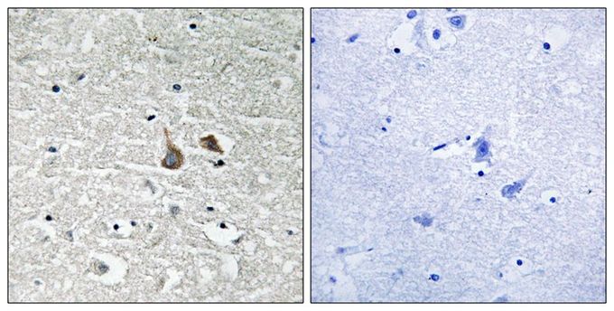
Immunohistochemical analysis of paraffin-embedded human brain tissue using IRAK1 (Phospho-Ser376) antibody (left) or the same antibody preincubated with blocking peptide (right).| Product Name | IRAK1 (phospho Ser376) antibody |
|---|---|
| Antibody Type | Primary Antibodies |
| Product description | Rabbit Polyclonal antibody to IRAK1 (phospho Ser376)This gene encodes the interleukin-1 receptor-associated kinase 1, one of two putative serine/threonine kinases that become associated with the interleukin-1 receptor (IL1R) upon stimulation. This gene is partially responsible for IL1-induced upregulation of the transcription factor NF-kappa B. Alternatively spliced transcript variants encoding different isoforms have been found for this gene. [provided by RefSeq, Jul 2008] |
| Immunogen | Peptide sequence around phosphorylation site of Serine 376(Q-S-S(p)-M-V) derived from Human IRAK1. |
| Modification | p-Ser376 |
| Clonality | Polyclonal |
|---|---|
| Isotype | IgG |
| Host Species | Rabbit |
| Tested Applications | IHC-P |
| IHC: 1:50 - 1:100: |
|
| Species Reactivity | Human |
| Concentration | 1mg/ml (Please refer to the vial label for the specific concentration) |
| Purification | Affinity purified |
| Gene Synonyms | IRAK IRAK1 pelle interleukin-1 receptor-associated kinase 1 |
|---|---|
| Target | IRAK1 (phospho Ser376) |
| Tissue Specificity | The antibody detects endogenous levels of IRAK1 only when phosphorylated at serine 376. |
Application
Immunohistochemical analysis of paraffin-embedded human brain tissue using IRAK1 (Phospho-Ser376) antibody (left) or the same antibody preincubated with blocking peptide (right).| Application Notes | IHC: 1:50 - 1:100: |
|---|
| Form | Liquid |
|---|---|
| Storage Instructions | Store at -20˚C/1 year |
| Storage Buffer | Rabbit IgG in phosphate buffered saline (without Mg2+ and Ca2+), pH 7.4, 150mM NaCl, 0.02% sodium azide and 50% glycerol. |


