Application
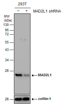
Non-transfected (–) and transfected (+) 293T whole cell extracts (30 µg) were separated by 12% SDS-PAGE, and the membrane was blotted with MAD2L1 antibody [C2C3], C-term diluted at 1:1000. The HRP-conjugated anti-rabbit IgG antibody was used to detect the primary antibody.Application
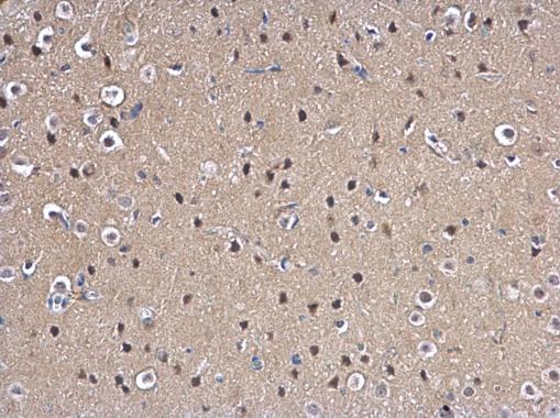
MAD2L1 antibody [C2C3], C-term detects MAD2L1 protein at nucleus on rat fore brain by immunohistochemical analysis. Sample: Paraffin-embedded rat fore brain. MAD2L1 antibody [C2C3], C-term dilution: 1:500.Application
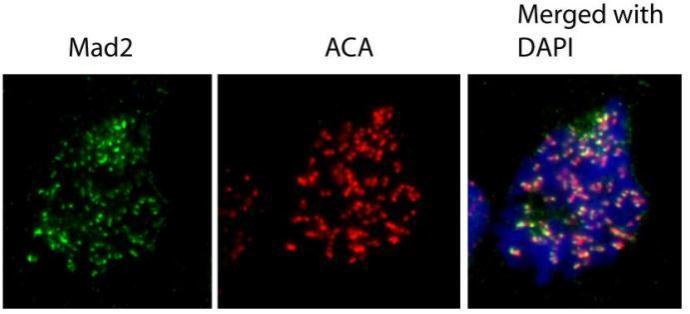
Immunofluorescence analysis of human osteosarcoma cell line U2OS, using MAD2L1 antibody at 1:1000 dilution.Application
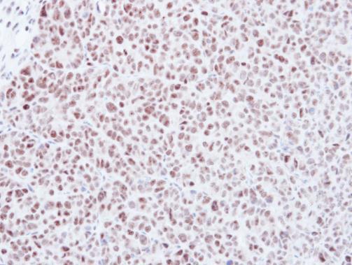
Immunohistochemical analysis of paraffin-embedded DLD-1 xenograft, using Mad2L1 antibody at 1:500 dilution.Application
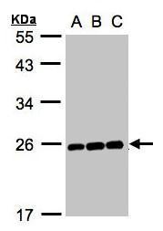
Sample (30 µg whole cell lysate) A:Hep G2 B:MOLT4 C:Raji 12% SDS PAGE diluted at 1:1000 The HRP-conjugated anti-rabbit IgG antibody was used to detect the primary antibody.Application
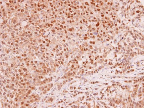
MAD2L1 antibody [C2C3], C-term detects MAD2L1 protein at cytoplasm and nucleus in human A549 xenograft by immunohistochemical analysis. Sample: Paraffin-embedded human A549 xenograft . MAD2L1 antibody [C2C3], C-term diluted at 1:250.Application
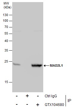
Immunoprecipitation of MAD2L1 protein from 293T whole cell extracts using 5 μg of MAD2L1 antibody [C2C3], C-term . Western blot analysis was performed using MAD2L1 antibody [C2C3], C-term . EasyBlot anti-Rabbit IgG was used as a secondary reagent.| Product Name | MAD2L1 antibody [C2C3], C-term |
|---|---|
| Antibody Type | Primary Antibodies |
| Product description | Rabbit Polyclonal antibody to MAD2L1 (MAD2 mitotic arrest deficient-like 1 (yeast))MAD2L1 is a component of the mitotic spindle assembly checkpoint that prevents the onset of anaphase until all chromosomes are properly aligned at the metaphase plate. MAD2L1 is related to the MAD2L2 gene located on chromosome 1. A MAD2 pseudogene has been mapped to chromosome 14. [provided by RefSeq] |
| Immunogen | Carrier-protein conjugated synthetic peptide encompassing a sequence within the C-terminus region of human MAD2L1. The exact sequence is proprietary. |
| Clonality | Polyclonal |
|---|---|
| Isotype | IgG |
| Host Species | Rabbit |
| Tested Applications | ICC/IFIHC-PIPWB |
| *Optimal dilutions/concentrations should be determined by the researcher. | |
| Species Reactivity | HumanRat |
| Predicted Reactivity | Rhesus Monkey, BovineRhesus Monkey (100%), Bovine (100%) |
| Concentration | 1mg/ml (Please refer to the vial label for the specific concentration) |
| Purification | Affinity purified |
| Gene Synonyms | HSMAD2 antibody, MAD2 antibody, MAD2L1 antibody, "MAD2 (mitotic arrest deficient, yeast, homolog)-like 1 antibody", MAD2-like protein 1 antibody, mitotic spindle assembly checkpoint protein MAD2A antibody, "mitotic arrest deficient, yeast, homolog-like 1 a |
|---|---|
| Molecular Weight(MW) | 24 kDa (note) |
Application
Non-transfected (–) and transfected (+) 293T whole cell extracts (30 µg) were separated by 12% SDS-PAGE, and the membrane was blotted with MAD2L1 antibody [C2C3], C-term diluted at 1:1000. The HRP-conjugated anti-rabbit IgG antibody was used to detect the primary antibody.
Application
MAD2L1 antibody [C2C3], C-term detects MAD2L1 protein at nucleus on rat fore brain by immunohistochemical analysis. Sample: Paraffin-embedded rat fore brain. MAD2L1 antibody [C2C3], C-term dilution: 1:500.
Application
Immunofluorescence analysis of human osteosarcoma cell line U2OS, using MAD2L1 antibody at 1:1000 dilution.
Application
Immunohistochemical analysis of paraffin-embedded DLD-1 xenograft, using Mad2L1 antibody at 1:500 dilution.
Application
Sample (30 µg whole cell lysate) A:Hep G2 B:MOLT4 C:Raji 12% SDS PAGE diluted at 1:1000 The HRP-conjugated anti-rabbit IgG antibody was used to detect the primary antibody.
Application
MAD2L1 antibody [C2C3], C-term detects MAD2L1 protein at cytoplasm and nucleus in human A549 xenograft by immunohistochemical analysis. Sample: Paraffin-embedded human A549 xenograft . MAD2L1 antibody [C2C3], C-term diluted at 1:250.
Application
Immunoprecipitation of MAD2L1 protein from 293T whole cell extracts using 5 μg of MAD2L1 antibody [C2C3], C-term . Western blot analysis was performed using MAD2L1 antibody [C2C3], C-term . EasyBlot anti-Rabbit IgG was used as a secondary reagent.| Application Notes | *Optimal dilutions/concentrations should be determined by the researcher. |
|---|
| Form | Liquid |
|---|---|
| Storage Instructions | Keep as concentrated solution. Aliquot and store at -20ºC or below. Avoid multiple freeze-thaw cycles. |
| Storage Buffer | 1XPBS, 40% Glycerol (pH7). 0.01% Thimerosal was added as a preservative. |


