Application
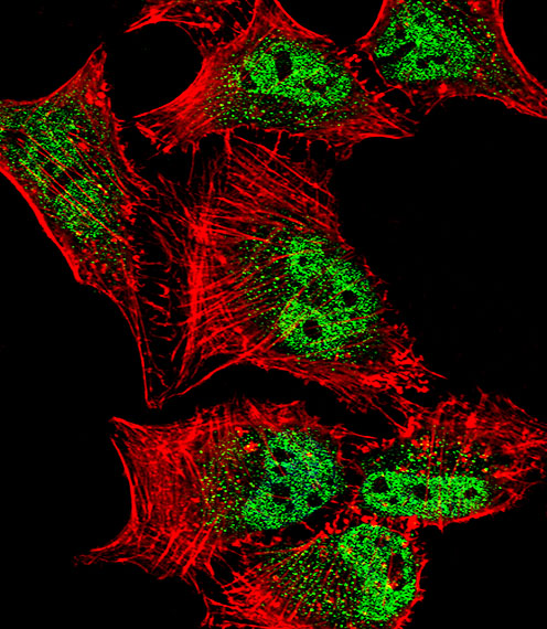
Fluorescent confocal image of Hela cell stained with MEF2C Antibody (S387).Hela cells were fixed with 4% PFA (20 min), permeabilized with Triton X-100 (0.1%, 10 min), then incubated with MEF2C primary antibody (1:25, 1 h at 37℃). For secondary antibody, Alexa Fluor® 488 conjugated donkey anti-rabbit antibody (green) was used (1:400, 50 min at 37℃).Cytoplasmic actin was counterstained with Alexa Fluor® 555 (red) conjugated Phalloidin (7units/ml, 1 h at 37℃). Nuclei were counterstained with DAPI (blue) (10 µg/ml, 10 min).MEF2C immunoreactivity is localized to vesicles and Nucleus significantly.Application
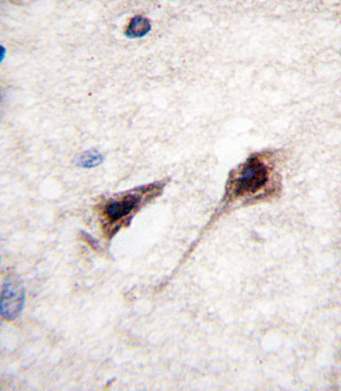
Formalin-fixed and paraffin-embedded human brain tissue reacted with MEF2C Antibody (S387) , which was peroxidase-conjugated to the secondary antibody, followed by DAB staining. This data demonstrates the use of this antibody for immunohistochemistry; clinical relevance has not been evaluated.Application
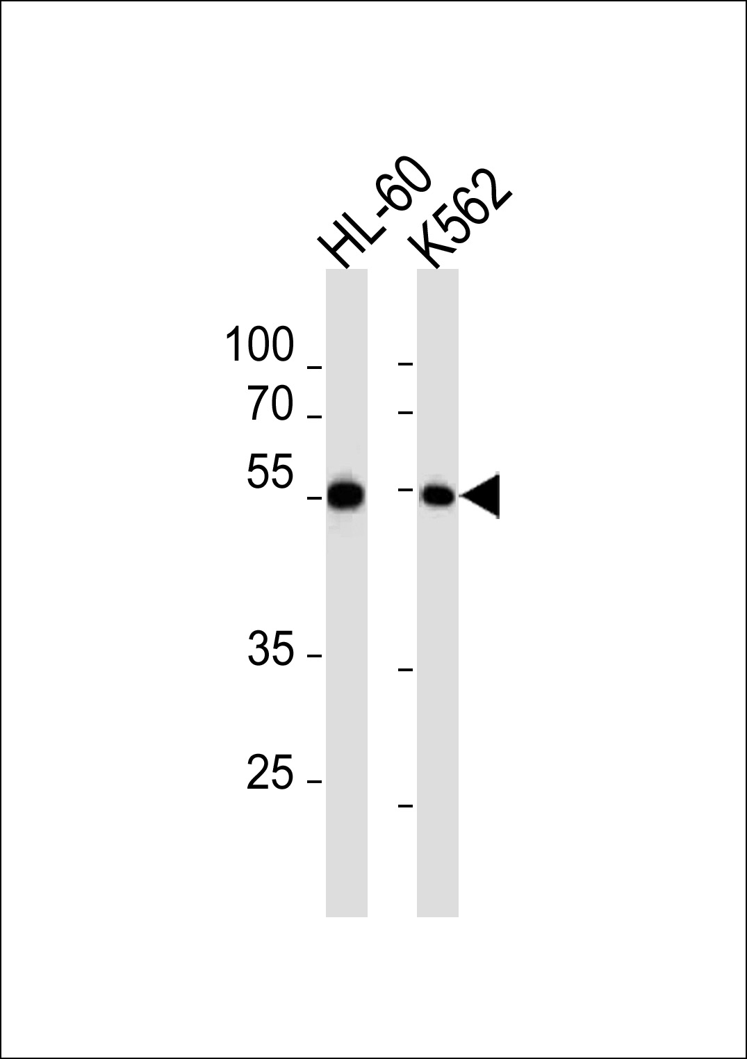
MEF2C Antibody (pS387) western blot analysis in HL-60,K562 cell line lysates (35ug/lane).This demonstrates the MEF2C antibody detected the MEF2C protein (arrow).| Product Name | MEF2C antibody |
|---|---|
| Antibody Type | Primary Antibodies |
| Product description | Rabbit polyclonal to MEF2C (S387)This locus encodes a member of the MADS box transcription enhancer factor 2 (MEF2) family of proteins, which play a role in myogenesis. The encoded protein, MEF2 polypeptide C, has both trans-activating and DNA binding activities. This protein may play a role in maintaining the differentiated state of muscle cells. Mutations and deletions at this locus have been associated with severe mental retardation, stereotypic movements, epilepsy, and cerebral malformation. Alternatively spliced transcript variants have been described. [provided by RefSeq] |
| Immunogen | KLH conjugated synthetic peptide corresponding to amino acid residues surrounding S387 of human MEF2C. |
| Clonality | Polyclonal |
|---|---|
| Isotype | IgG |
| Host Species | Rabbit |
| Tested Applications | ICC/IFIHC-PWB |
| ELISA: Use at 1:1,000: |
|
| Species Reactivity | Human |
| Concentration | 0.2mg/ml |
| Purification | Affinity purified |
| Gene Synonyms | Myocyte-specific enhancer factor 2C |
|---|---|
| Target | MEF2C |
| Molecular Weight(MW) | 51.09 kDa (note) |
Application
Fluorescent confocal image of Hela cell stained with MEF2C Antibody (S387).Hela cells were fixed with 4% PFA (20 min), permeabilized with Triton X-100 (0.1%, 10 min), then incubated with MEF2C primary antibody (1:25, 1 h at 37℃). For secondary antibody, Alexa Fluor® 488 conjugated donkey anti-rabbit antibody (green) was used (1:400, 50 min at 37℃).Cytoplasmic actin was counterstained with Alexa Fluor® 555 (red) conjugated Phalloidin (7units/ml, 1 h at 37℃). Nuclei were counterstained with DAPI (blue) (10 µg/ml, 10 min).MEF2C immunoreactivity is localized to vesicles and Nucleus significantly.
Application
Formalin-fixed and paraffin-embedded human brain tissue reacted with MEF2C Antibody (S387) , which was peroxidase-conjugated to the secondary antibody, followed by DAB staining. This data demonstrates the use of this antibody for immunohistochemistry; clinical relevance has not been evaluated.
Application
MEF2C Antibody (pS387) western blot analysis in HL-60,K562 cell line lysates (35ug/lane).This demonstrates the MEF2C antibody detected the MEF2C protein (arrow).| Application Notes | ELISA: Use at 1:1,000: |
|---|
| Form | Liquid |
|---|---|
| Storage Instructions | Store at at 2-8°C for up to 6 months. For long term storage, aliquot and store at -20 °C. Avoid repeated freeze/thaw cycles. |
| Storage Buffer | PBS with 0.09% (W/V) sodium azide |


