Application
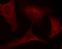
Immunofluorescence staining of methanol-fixed Hela cells using MEK1 (Phospho-Ser221) Antibody .Application
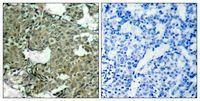
Immunohistochemical analysis of paraffin-embedded human breast carcinoma tissue using MEK1 (Phospho-Ser221) Antibody (left) or the same antibody preincubated with blocking peptide (right).Application
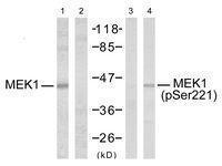
Western blot analysis of extracts from Hela cells untreated or treated with EGF using MEK1 (Phospho-Ser221) Antibody .Application
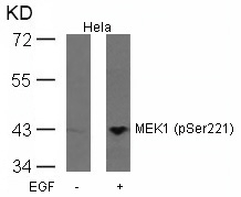
Western blot analysis of extracts from Hela cells untreated or treated with EGF using MEK1(Phospho-Ser221)IHC
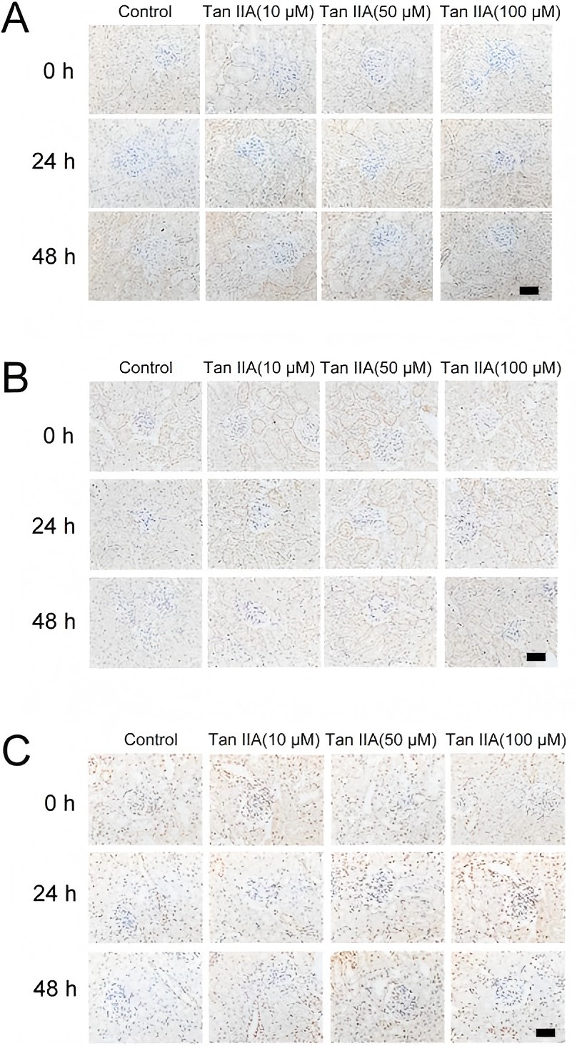
Tan IIA increased the expression of p-MEK (A), p-ERK1/2 (B) and p-GSK-3β (C), as assessed by immunohistochemical analysis.WB
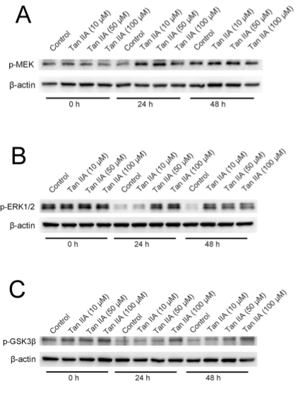
Tan IIA increases the expression of p-MEK (A), p-ERK1/2 (B) and p-GSK-3β (C), as detected by immunoblotting.WB
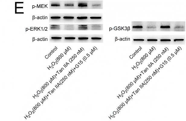
(E) Tan IIA increased the expression of p-MEK, p-ERK1/2 and p-GSK-3β, as detected by immunoblotting. G15 could diminish the expression of these proteins induced by Tan IIA.| Product Name | MEK1 (phospho Ser221) antibody |
|---|---|
| Antibody Type | Primary Antibodies |
| Product description | Rabbit Polyclonal antibody to MEK1 (Phospho Ser221)Catalyzes the concomitant phosphorylation of a threonine and a tyrosine residue in a Thr-Glu-Tyr sequence located in MAP kinases. Activates ERK1 and ERK2 MAP kinases. |
| Immunogen | Peptide sequence around phosphorylation site of serine 221 (A-N-S(p)-F-V) derived from Human MEK1. |
| Modification | p-Ser221 |
| Clonality | Polyclonal |
|---|---|
| Host Species | Rabbit |
| Tested Applications | ICC/IFIHC-PWB |
| Suggested starting dilutions are as follows. WB: 1:500~1:1000, IHC-P: 1:50~1:100, ICC/IF: 1:100~1:200. Not yet tested in other applications. Optimal working dilutions should be determined experimentally by the end user.: |
|
| Species Reactivity | HumanMouseRat |
| Concentration | 1mg/ |
| Purification | Affinity purified |
| Gene Synonyms | MAPKK1 MEK1 MKK1 PRKMK1 MAP2K1 protein kinase mitogen-activated kinase 1 (MAP kinase kinase 1) ERK activator kinase 1 MEK 1 MAP kinase kinase 1 MAPK/ERK kinase 1 dual specificity mitogen-activated protein kinase kinase 1 MAPKK 1 mitogen-activ |
|---|---|
| Target | MEK1 (Phospho Ser221) |
| Molecular Weight(MW) | 45 (note) |
Application
Immunofluorescence staining of methanol-fixed Hela cells using MEK1 (Phospho-Ser221) Antibody .
Application
Immunohistochemical analysis of paraffin-embedded human breast carcinoma tissue using MEK1 (Phospho-Ser221) Antibody (left) or the same antibody preincubated with blocking peptide (right).
Application
Western blot analysis of extracts from Hela cells untreated or treated with EGF using MEK1 (Phospho-Ser221) Antibody .
Application
Western blot analysis of extracts from Hela cells untreated or treated with EGF using MEK1(Phospho-Ser221)
IHC
Tan IIA increased the expression of p-MEK (A), p-ERK1/2 (B) and p-GSK-3β (C), as assessed by immunohistochemical analysis.
WB
Tan IIA increases the expression of p-MEK (A), p-ERK1/2 (B) and p-GSK-3β (C), as detected by immunoblotting.
WB
(E) Tan IIA increased the expression of p-MEK, p-ERK1/2 and p-GSK-3β, as detected by immunoblotting. G15 could diminish the expression of these proteins induced by Tan IIA.| Application Notes | Suggested starting dilutions are as follows. WB: 1:500~1:1000, IHC-P: 1:50~1:100, ICC/IF: 1:100~1:200. Not yet tested in other applications. Optimal working dilutions should be determined experimentally by the end user.: |
|---|
| Form | Liquid |
|---|---|
| Storage Instructions | Keep as concentrated solution. Aliquot and store at -20ºC or below. Avoid multiple freeze-thaw cycles. |
| Storage Buffer | saline (without Mg2+ and Ca2+), pH 7.4, 150mM NaCl, 0.02% sodium azide and 50% glycerol. |


