Application
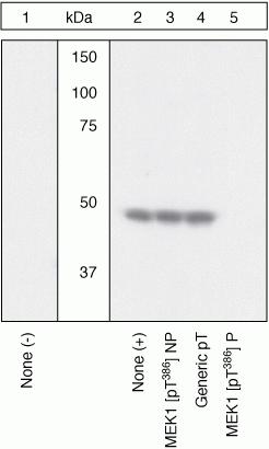
Extracts of NIH3T3 cells untreated (1) or treated with 50ng/mL PDGF for 15 minutes (2-5) were resolved by SDS-PAGE on a 10% Tris-glycine gel and transferred to PVDF. The membrane was blocked with a 4% BSA-TBST buffer for one hour at room temperature, then incubated with the MEK1 [pT386] antibody in a 1% BSA-TBST buffer for two hours at room temperature, following prior incubation with: no peptide (1, 2), the non-phosphopeptide corresponding to the phosphopeptide immunogen (3), a generic phosphothreonine-containing peptide (4), or the phosphopeptide immunogen (5). After washing, the membrane was incubated with goat F(ab)2 anti-rabbit IgG HRP conjugate and signals were detected.The data show that only the phosphopeptide corresponding to MEK1 [pT386] blocks the antibody signal, demonstrating the specificity of the antibody. The data also show the upregulation of MEK1 [pT386] upon PDGF treatment in this cell system.| Product Name | MEK1 (phospho Thr386) antibody |
|---|---|
| Antibody Type | Primary Antibodies |
| Product description | Rabbit polyclonal to MEK1 ( phospho T386 )Mitogen activated protein kinase kinase 2 (MEK 2 or MAPKK 2) is a member of a family of tyrosine/threonine protein kinases that activate the ERK 1 and 2/MAPK enzymes by phosphorylating both residues within the threonine - glutamate - tyrosine (TEY) motif in the activation loop. MEK 1 + 2 are also activated by dual phosphorylation, which occurs on serine 218 and 222, in the activation loop of the MEK. Threonine 386 of MEK 1 is phosphorylated by ERK 2, which serves as a negative feedback loop by suppressing activation of MEK 1. |
| Immunogen | Phosphopeptide derived from a region of human MEK 1 that contains threonine 386. |
| Modification | p-Thr386 |
| Clonality | Polyclonal |
|---|---|
| Isotype | IgG |
| Host Species | Rabbit |
| Tested Applications | WB |
| Recommended dilutions: WB: 1/1000. Detects a band of approximately 50 kDa. Not yet tested in other applications. Optimal dilutions/concentrations should be determined by the end user.: |
|
| Species Reactivity | HumanMouseRat |
| Concentration | 1mg/ml |
| Purification | Affinity purified |
| Gene Synonyms | MAPKK1 MEK1 MKK1 PRKMK1 MEK1 (phospho T386) MEK1 (phospho Thr386) MEK1 (pT386) MEK1 (pThr386) MEK1 phospho T386 MEK1 phospho Thr386 Phospho MEK1 (T386) Phospho MEK1 (Thr386) Phospho MEK1 (pT386) Phospho MEK1 (pThr386) |
|---|---|
| Target | MEK1 |
| Cellular Localization | Cytoplasmic |
Application
Extracts of NIH3T3 cells untreated (1) or treated with 50ng/mL PDGF for 15 minutes (2-5) were resolved by SDS-PAGE on a 10% Tris-glycine gel and transferred to PVDF. The membrane was blocked with a 4% BSA-TBST buffer for one hour at room temperature, then incubated with the MEK1 [pT386] antibody in a 1% BSA-TBST buffer for two hours at room temperature, following prior incubation with: no peptide (1, 2), the non-phosphopeptide corresponding to the phosphopeptide immunogen (3), a generic phosphothreonine-containing peptide (4), or the phosphopeptide immunogen (5). After washing, the membrane was incubated with goat F(ab)2 anti-rabbit IgG HRP conjugate and signals were detected.The data show that only the phosphopeptide corresponding to MEK1 [pT386] blocks the antibody signal, demonstrating the specificity of the antibody. The data also show the upregulation of MEK1 [pT386] upon PDGF treatment in this cell system.| Positive Control | NIH3T3 cells +/- PDGF, A431 +/- EGF, PC12 +/- NGF |
|---|---|
| Application Notes | Recommended dilutions: WB: 1/1000. Detects a band of approximately 50 kDa. Not yet tested in other applications. Optimal dilutions/concentrations should be determined by the end user.: |
| Form | Liquid |
|---|---|
| Storage Instructions | Upon receipt - Keep as concentrated solution. Aliquot and store at -20ºC or below. Avoid freeze-thaw cycles. |
| Storage Buffer | Preservative: 0.05% Sodium Azide; Constituents: PBS, 50% glycerol, 1mg/ml BSA (IgG, protease free). pH 7.3 |


