Application
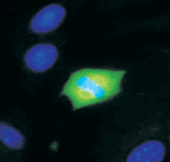
Mouse embryonic fibroblasts stained with anti-MEK1⁄2 [pS222] . The cell is actively dividing. Blue represents chromosomes in anaphase of mitotic cell division. Green shows mitotic spindle expressing phosphorylated MEK1⁄2.Application
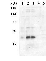
Extracts prepared from NIH3T3 cells left untreated (lane 1) or treated (lanes 2-5) with PDGF were resolved by SDS-PAGE on a 10% Tris-glycine gel and transferred to nitrocellulose. Membranes were blocked with a 5% BSA-TBST buffer overnight at 4 degree C, then incubated with MEK1 +MEK2 antibody.Application
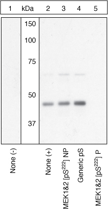
Extracts of NIH3T3 cells untreated (lane 1) or treated with 50 ng/mL PDGF for 15 minutes (2-5) were resolved by SDS-PAGE on a 10% Tris-glycine gel and transferred to PVDF. The membrane was blocked with a 4% BSA-TBST buffer overnight at 4°C, then incubated with the MEK1&2 [pS222] antibody for two hours at room temperature in a 1% BSA-TBST buffer, following prior incubation with: no peptide (1, 2), the non-phosphopeptide corresponding to the phosphopeptide immunogen (3), a generic phosphoserine-containing peptide (4), or the phosphopeptide immunogen (5). After washing, the membrane was incubated with goat F(ab)2 anti-rabbit IgG alkaline phosphatase and signals were detected. The data show that only the phosphopeptide corresponding to MEK1&2 [pS222] block the antibody signal, demonstrating the specificity of the antibody. The data also show the induction of MEK1&2 [pS222] phosphorylation by the addition of PDGF to this cell system.| Product Name | MEK1 + MEK2 (phospho Ser222) antibody |
|---|---|
| Antibody Type | Primary Antibodies |
| Product description | Rabbit polyclonal to MEK1 + MEK2 ( phospho S222 )Microtubule-associated protein kinase kinase 1 + 2 (MEK 1 + 2 or MAPKK 1+ 2) are members of a tyrosine/threonine protein kinase family that activate the ERK 1 + 2/MAPK enzymes by phosphorylating both residues within the threonine - glutamic acid - tyrosine (TEY) motif in the activation loop. MEK 1 + 2 are also activated by dual-phosphorylation, which occurs on serines 218 and 222, in the activation loop of the MEKs. |
| Immunogen | Synthetic peptide (Human) derived from a region of human MEK 1 + 2 that contains serine 222. The sequence is conserved in many species including mouse, rat, chimp, chicken, frog, and others. |
| Modification | p-Ser222 |
| Clonality | Polyclonal |
|---|---|
| Isotype | IgG |
| Host Species | Rabbit |
| Tested Applications | ICC/IFWB |
| WB: Use at a concentration of 0.1 - 1.0 μg/ml. Optimal dilutions/concentrations should be determined by the end user.: |
|
| Species Reactivity | HumanMouseRatChimpanzeeXenopus |
| Concentration | 0.5mg/ml |
| Purification | Affinity purified |
| Gene Synonyms | MAPKK1 FLJ26075 MEK1 MAPKK2 MKK1 MEK2 PRKMK1 MKK2 PRKMK2 MEK1 + MEK2 (phospho S222) MEK1 + MEK2 (phospho Ser222) MEK1 + MEK2 (pS222) MEK1 + MEK2 (pSer222) MEK1 + MEK2 phospho S222 MEK1 + MEK2 phospho Ser222 Phospho MEK1 + MEK2 (S222) Phosp |
|---|---|
| Target | MEK1 + MEK2 phospho S222 |
Application
Mouse embryonic fibroblasts stained with anti-MEK1⁄2 [pS222] . The cell is actively dividing. Blue represents chromosomes in anaphase of mitotic cell division. Green shows mitotic spindle expressing phosphorylated MEK1⁄2.
Application
Extracts prepared from NIH3T3 cells left untreated (lane 1) or treated (lanes 2-5) with PDGF were resolved by SDS-PAGE on a 10% Tris-glycine gel and transferred to nitrocellulose. Membranes were blocked with a 5% BSA-TBST buffer overnight at 4 degree C, then incubated with MEK1 +MEK2 antibody.
Application
Extracts of NIH3T3 cells untreated (lane 1) or treated with 50 ng/mL PDGF for 15 minutes (2-5) were resolved by SDS-PAGE on a 10% Tris-glycine gel and transferred to PVDF. The membrane was blocked with a 4% BSA-TBST buffer overnight at 4°C, then incubated with the MEK1&2 [pS222] antibody for two hours at room temperature in a 1% BSA-TBST buffer, following prior incubation with: no peptide (1, 2), the non-phosphopeptide corresponding to the phosphopeptide immunogen (3), a generic phosphoserine-containing peptide (4), or the phosphopeptide immunogen (5). After washing, the membrane was incubated with goat F(ab)2 anti-rabbit IgG alkaline phosphatase and signals were detected. The data show that only the phosphopeptide corresponding to MEK1&2 [pS222] block the antibody signal, demonstrating the specificity of the antibody. The data also show the induction of MEK1&2 [pS222] phosphorylation by the addition of PDGF to this cell system.| Positive Control | NIH3T3 cells +/- PDGF, A431 +/- EGF, PC12 +/- NGF |
|---|---|
| Application Notes | WB: Use at a concentration of 0.1 - 1.0 μg/ml. Optimal dilutions/concentrations should be determined by the end user.: |
| Form | Liquid |
|---|---|
| Storage Instructions | For short-term storage, store at 4° C. For long-term storage, aliquot and store at -20ºC or below. Avoid multiple freeze-thaw cycles. |
| Storage Buffer | Preservative: 0.05% Sodium Azide; Constituents: PBS, 1.0mg/ml BSA (IgG, protease free). pH 7.4 |


