Application
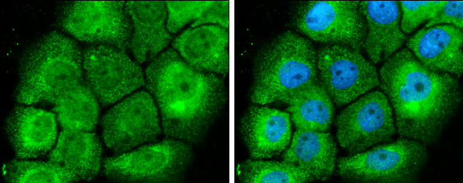
MEK1+2 antibody detects MEK1+2 protein at cytoplasm and nucleus by immunofluorescent analysis. Sample: A431 cells were fixed in 4% paraformaldehyde at RT for 15 min. Green: MEK1+2 protein stained by MEK1+2 antibody diluted at 1:500. Blue: Hoechst 33342 staining.Application
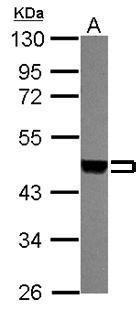
Sample (30 ug of whole cell lysate) A: 293T 10% SDS PAGE diluted at 1:3000Application
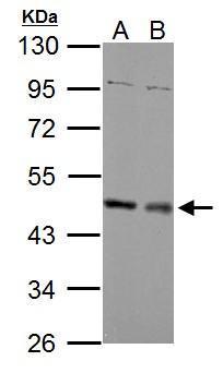
Sample (30 ug of whole cell lysate) A: NIH-3T3 B: JC 10% SDS PAGE diluted at 1:2000Application
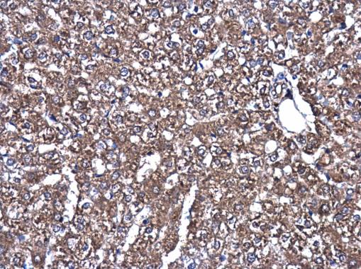
MEK1+2 antibody detects MEK1+2 protein at cytoplasm in rat liver by immunohistochemical analysis. Sample: Paraffin-embedded rat liver. MEK1+2 antibody diluted at 1:500.| Product Name | MEK1+2 antibody |
|---|---|
| Antibody Type | Primary Antibodies |
| Product description | Rabbit Polyclonal antibody to MEK1+2 (mitogen-activated protein kinase kinase 1)The protein encoded by this gene is a member of the dual specificity protein kinase family, which acts as a mitogen-activated protein (MAP) kinase kinase. MAP kinases, also known as extracellular signal-regulated kinases (ERKs), act as an integration point for multiple biochemical signals. This protein kinase lies upstream of MAP kinases and stimulates the enzymatic activity of MAP kinases upon wide variety of extra- and intracellular signals. As an essential component of MAP kinase signal transduction pathway, this kinase is involved in many cellular processes such as proliferation, differentiation, transcription regulation and development. [provided by RefSeq] |
| Immunogen | Recombinant protein encompassing a sequence within the center region of human MEK1+2. The exact sequence is proprietary. |
| Clonality | Polyclonal |
|---|---|
| Isotype | IgG |
| Host Species | Rabbit |
| Tested Applications | ICC/IFIHC-PWB |
| *Optimal dilutions/concentrations should be determined by the researcher.
: |
|
| Species Reactivity | HumanMouseRat |
| Predicted Reactivity | Zebrafish, Xenopus laevis, Dog, Pig, Rabbit, Chicken, Rhesus Monkey, Chimpanzee, BovineZebrafish (94%), Xenopus laevis (96%), Dog (100%), Pig (96%), Rabbit (100%), Chicken (98%), Rhesus Monkey (100%), Chimpanzee (98%), Bovine (100%) |
| Concentration | 1mg/ml (Please refer to the vial label for the specific concentration) |
| Purification | Affinity purified |
| Gene Synonyms | MAPKK1 antibody MEK1 antibody MKK1 antibody PRKMK1 antibody MAP2K1 antibody "protein kinase mitogen-activated kinase 1 (MAP kinase kinase 1) antibody" ERK activator kinase 1 antibody MEK 1 antibody MAP kinase kinase 1 antibody MAPK/ERK kinase 1 |
|---|---|
| Molecular Weight(MW) | 43 kDa (note) (2) |
Application
MEK1+2 antibody detects MEK1+2 protein at cytoplasm and nucleus by immunofluorescent analysis. Sample: A431 cells were fixed in 4% paraformaldehyde at RT for 15 min. Green: MEK1+2 protein stained by MEK1+2 antibody diluted at 1:500. Blue: Hoechst 33342 staining.
Application
Sample (30 ug of whole cell lysate) A: 293T 10% SDS PAGE diluted at 1:3000
Application
Sample (30 ug of whole cell lysate) A: NIH-3T3 B: JC 10% SDS PAGE diluted at 1:2000
Application
MEK1+2 antibody detects MEK1+2 protein at cytoplasm in rat liver by immunohistochemical analysis. Sample: Paraffin-embedded rat liver. MEK1+2 antibody diluted at 1:500.| Positive Control | 293T , NIH-3T3 , JC |
|---|---|
| Application Notes | *Optimal dilutions/concentrations should be determined by the researcher.
: |
| Form | Liquid |
|---|---|
| Storage Instructions | Keep as concentrated solution. Aliquot and store at -20ºC or below. Avoid multiple freeze-thaw cycles. |
| Storage Buffer | 1XPBS, 1% BSA, 20% Glycerol (pH7). 0.01% Thimerosal was added as a preservative. |


