Application
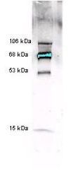
Anti-Myeloperoxidase [Human Leukocytes] detects multiple MPO subunits and chain combinations by western blot. Polyclonal rabbit-anti-Myeloperoxidase was used at a 1:5000 dilution to detect 1.0 ug of human myeloperoxidase. This antibody detects a multiple bands corresponding to 53 kDa and 15 kDa polypeptides and chain combinations forming 68 kDa and 106 kDa proteins. The staining of the 68 kDa band is so intense that is over saturates the signal detection. A 4-20% gradient gel was used to separate the protein by SDS-PAGE. The protein was transferred to nitrocellulose using standard methods. After blocking the membrane was probed with the primary antibody for 2 h at room temperature followed by washes and reaction with a 1:5,000 dilution of IRDye™800 conjugated Goat anti Rabbit IgG [H&L] for 30 min at room temperature.| Product Name | Myeloperoxidase antibody |
|---|---|
| Antibody Type | Primary Antibodies |
| Product description | Rabbit Polyclonal antibody to MyeloperoxidaseHuman myeloperoxidase (MPO) is a dimeric protein composed of two heavy subunits (53 kDa) and two light subunits (15 kDa). Each MPO molecule contains two prosthetic porphyrins which play an important role in the catalytic cycle. Molecular weights for MPO isoforms from pools of normal human samples range from 114,000 to 140,000 daltons reflecting a heterogeneous mixture of isoforms when assayed under non-reducing conditions of SDS-PAGE. Often MPO from a single donor will yield a homogenous preparation reflecting a single isoform. The carbohydrate component of MPO, consisting of mannose, glucose and N-acetylglucosamine residues is 2.5%. MPO is inhibited by azide and other compounds. MPO is stored in primary granules of neutrophils and serves as a bactericidal agent in that MPO catalyzes the production of hypochlorous acid (HOCl), a powerful oxidant. HOCl is derived from chloride ion (Cl-) and hydrogen peroxide (H2O2). In a number of inflammatory situations, MPO is released into the extracellular matrix where its measurement can be used as an indication of neutrophil activation. |
| Immunogen | Myeloperoxidase [Human Leukocytes] |
| Clonality | Polyclonal |
|---|---|
| Host Species | Rabbit |
| Tested Applications | ELISAIPWB |
| *Optimal dilutions/concentrations should be determined by the researcher.
: |
|
| Species Reactivity | Human |
| Concentration | 1mg/ml |
| Purification | Affinity purified |
| Gene Synonyms | MPO |
|---|---|
| Target | Myeloperoxidase |
| Cellular Localization | Lysosomal |
Application
Anti-Myeloperoxidase [Human Leukocytes] detects multiple MPO subunits and chain combinations by western blot. Polyclonal rabbit-anti-Myeloperoxidase was used at a 1:5000 dilution to detect 1.0 ug of human myeloperoxidase. This antibody detects a multiple bands corresponding to 53 kDa and 15 kDa polypeptides and chain combinations forming 68 kDa and 106 kDa proteins. The staining of the 68 kDa band is so intense that is over saturates the signal detection. A 4-20% gradient gel was used to separate the protein by SDS-PAGE. The protein was transferred to nitrocellulose using standard methods. After blocking the membrane was probed with the primary antibody for 2 h at room temperature followed by washes and reaction with a 1:5,000 dilution of IRDye™800 conjugated Goat anti Rabbit IgG [H&L] for 30 min at room temperature.| Application Notes | *Optimal dilutions/concentrations should be determined by the researcher.
: |
|---|
| Form | Liquid |
|---|---|
| Storage Instructions | Keep as concentrated solution. Aliquot and store at -20oC or below. Avoid multiple freeze-thaw cycles. |
| Storage Buffer | 0.02 M Potassium Phosphate, 0.15 M Sodium Chloride, pH 7.2 and 0.01% (w/v) Sodium Azide |


