Application
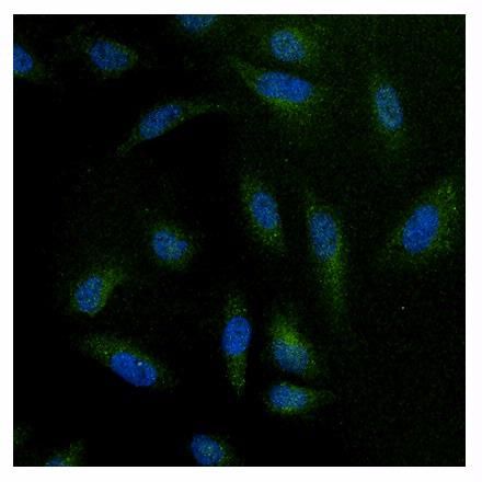
Immunofluorescent analysis of NFAT3 in HeLa cells. Formalin-fixed cells were permeabilized with 0.1% Triton X-100 in TBS for 10 minutes at room temperature. Cells were then blocked with 1% BSA for 15 minutes at room temperature. Cells were probed with NFAT3 antibody at a dilution of 1:100 for at least 1 hour at room temperature. Cells were washed with PBS and incubated with a proper secondary antibody . Nuclei (blue) were stained with Hoechst 33342 dye. Images were taken at 20X magnification.Application
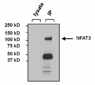
Immunoprecipitation of NFAT3 from MCF7 cell lysate. The antigen-antibody complex was formed by incubating 500μg whole cell lysate with 3μg of NFAT3 antibody overnight on a rocking platform at 4°C. The immune-complex was captured on 50μl Protein A/G Agarose. Captured immune-complexes were washed and eluted. Samples were resolved on a 4-20% Tris-HCl polyacrylamide gel. Proteins were transferred to PVDF membrane and blocked with 5% Milk/TBS-0.1%Tween for at least 1 hour. Membranes were washed in TBS-0.1%Tween 20 and probed with NFAT3 antibody and a proper secondary antibody. Membranes were washed and chemiluminescent detection performed.Application
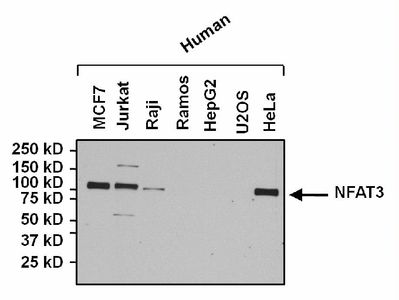
Western blot analysis of NFAT3 in 25ug of various whole cell lysates. Proteins were transferred to a PVDF membrane and blocked with 5% Milk/TBST for at least 1 hour. Membranes were probed with NFAT3 antibody at a dilution of 1:1000 overnight at 4°C on a rocking platform. Membranes were washed in TBS-0.1%Tween 20 and probed with a proper secondary. Membranes were washed and chemiluminescent detection performed.| Product Name | NFAT3 antibody |
|---|---|
| Antibody Type | Primary Antibodies |
| Product description | Rabbit polyclonal antibody to NFAT3The product of this gene is a member of the nuclear factors of activated T cells DNA-binding transcription complex. This complex consists of at least two components: a preexisting cytosolic component that translocates to the nucleus upon T cell receptor (TCR) stimulation and an inducible nuclear component. Other members of this family of nuclear factors of activated T cells also participate in the formation of this complex. The product of this gene plays a role in the inducible expression of cytokine genes in T cells, especially in the induction of the IL-2 and IL-4. Alternatively spliced transcript variants encoding different isoforms have been noted for this gene. [provided by RefSeq, Oct 2008] |
| Immunogen | Synthetic peptide corresponding to residues C R(887) D L S G F P A P P G E E P P A(902) of human NFAT3. |
| Clonality | Polyclonal |
|---|---|
| Isotype | IgG |
| Host Species | Rabbit |
| Tested Applications | ICC/IFIHCIPWB |
| ICC/IF: 1:100: |
|
| Species Reactivity | HumanMouse |
| Concentration | 1mg/ml (Please refer to the vial label for the specific concentration) |
| Purification | Affinity purified |
| Gene Synonyms | nuclear factor of activated T-cells cytoplasmic calcineurin-dependent 4 NFATC4 NF-ATc4 |
|---|---|
| Target | NFAT3 |
Application
Immunofluorescent analysis of NFAT3 in HeLa cells. Formalin-fixed cells were permeabilized with 0.1% Triton X-100 in TBS for 10 minutes at room temperature. Cells were then blocked with 1% BSA for 15 minutes at room temperature. Cells were probed with NFAT3 antibody at a dilution of 1:100 for at least 1 hour at room temperature. Cells were washed with PBS and incubated with a proper secondary antibody . Nuclei (blue) were stained with Hoechst 33342 dye. Images were taken at 20X magnification.
Application
Immunoprecipitation of NFAT3 from MCF7 cell lysate. The antigen-antibody complex was formed by incubating 500μg whole cell lysate with 3μg of NFAT3 antibody overnight on a rocking platform at 4°C. The immune-complex was captured on 50μl Protein A/G Agarose. Captured immune-complexes were washed and eluted. Samples were resolved on a 4-20% Tris-HCl polyacrylamide gel. Proteins were transferred to PVDF membrane and blocked with 5% Milk/TBS-0.1%Tween for at least 1 hour. Membranes were washed in TBS-0.1%Tween 20 and probed with NFAT3 antibody and a proper secondary antibody. Membranes were washed and chemiluminescent detection performed.
Application
Western blot analysis of NFAT3 in 25ug of various whole cell lysates. Proteins were transferred to a PVDF membrane and blocked with 5% Milk/TBST for at least 1 hour. Membranes were probed with NFAT3 antibody at a dilution of 1:1000 overnight at 4°C on a rocking platform. Membranes were washed in TBS-0.1%Tween 20 and probed with a proper secondary. Membranes were washed and chemiluminescent detection performed.| Application Notes | ICC/IF: 1:100: |
|---|
| Form | Liquid |
|---|---|
| Storage Instructions | -20° C, Avoid Freeze/Thaw Cycles |
| Storage Buffer | PBS containing 0.05 % sodium azide and 1 mg/ml BSA |


