Application
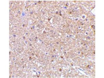
Immunohistochemistry showing detection of NogoA in mouse brain tissue. Primary antibody was used at 2.5 µg/ml.Application
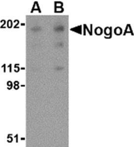
Western blot demonstrating detection of NogoA in human brain tissue lysate. Primary antibody was used at concentration of 0.5 µg/mL (A) and 1.0 µg/mL (B).| Product Name | Nogo A antibody |
|---|---|
| Antibody Type | Primary Antibodies |
| Product description | Rabbit Polyclonal antibody to Nogo ANogoA is a member of a family of integral membrane proteins termed reticulons that are thought to be involved in numerous disorders including neurodegenerative diseases. Reticulon proteins are known to regulate many cellular processes and interact with multiple proteins and receptors such as BACE. NogoA was initially identified as a myelin-associated neurite outgrowth inhibitor. It is highly expressed in oligodendrocytes in the white matter of the CNS; blocking its activity with antibodies or other factors results in improved axon regrowth and functional recovery in experimental CNS lesion models. NogoA has also been suggested to play a role in neurodegenerative diseases such as Amyotrophic lateral sclerosis, in which case NogoA is found at elevated levels in postmortem muscular samples, and multiple sclerosis (MS), in which case autoantibodies to NogoA have been found in serum and cerebrospinal fluid in MS patients. |
| Immunogen | This affinity purified antibody was prepared from whole rabbit serum produced by repeated immunizations with a synthetic peptide corresponding to an internal region of human Nogo A protein. |
| Clonality | Polyclonal |
|---|---|
| Isotype | IgG |
| Host Species | Rabbit |
| Tested Applications | ELISAIHCWB |
| *Optimal dilutions/concentrations should be determined by the researcher.
: |
|
| Species Reactivity | HumanMouseRat |
| Concentration | 1mg/ml |
| Purification | Affinity purified |
| Gene Synonyms | RTN4B2 RTN4B1 RTN4A RTN X RTN4C NI220/250 ASY NBLA10545 NOGO B RTN4 NSP NOGO NOGOC NOGO A NBLA00271 A |
|---|---|
| Target | Nogo A |
| Tissue Specificity | At least five isoforms of Nogo are known to exist; this antibody is specific for Nogo A and Nogo E. |
| Cellular Localization | Anchored to the membrane of the endoplasmic reticulum through 2 putative transmembrane domains. |
Application
Immunohistochemistry showing detection of NogoA in mouse brain tissue. Primary antibody was used at 2.5 µg/ml.
Application
Western blot demonstrating detection of NogoA in human brain tissue lysate. Primary antibody was used at concentration of 0.5 µg/mL (A) and 1.0 µg/mL (B).| Application Notes | *Optimal dilutions/concentrations should be determined by the researcher.
: |
|---|
| Form | Liquid |
|---|---|
| Storage Instructions | Store vial at -20° C prior to opening. Aliquot contents and freeze at -20° C or below for extended storage. Avoid cycles of freezing and thawing. |
| Storage Buffer | 0.02 M Potassium Phosphate, 0.15 M Sodium Chloride, pH 7.2 and 0.01% (w/v) Sodium Azide |


