Application
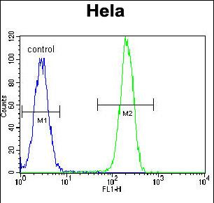
NPM1 Antibody (C-term) flow cytometric analysis of Hela cells (right histogram) compared to a negative control cell (left histogram).FITC-conjugated goat-anti-rabbit secondary antibodies were used for the analysis.Application
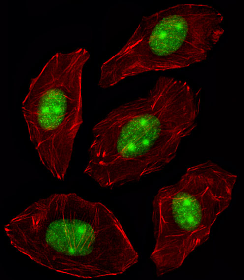
Fluorescent image of A549 cell stained with NPM1 Antibody (C-term).A549 cells were fixed with 4% PFA (20 min), permeabilized with Triton X-100 (0.1%, 10 min), then incubated with NPM1 primary antibody (1:25, 1 h at 37℃). For secondary antibody, Alexa Fluor® 488 conjugated donkey anti-rabbit antibody (green) was used (1:400, 50 min at 37℃).Cytoplasmic actin was counterstained with Alexa Fluor® 555 (red) conjugated Phalloidin (7units/ml, 1 h at 37℃).NPM1 immunoreactivity is localized to Nucleus and Nucleolus significantly.Application
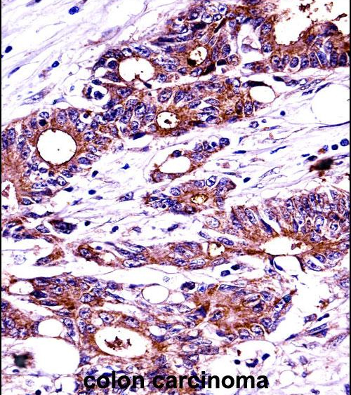
NPM1 Antibody (C-term) immunohistochemistry analysis in formalin fixed and paraffin embedded human colon carcinoma followed by peroxidase conjugation of the secondary antibody and DAB staining.This data demonstrates the use of NPM1 Antibody (C-term) for immunohistochemistry. Clinical relevance has not been evaluated.Application
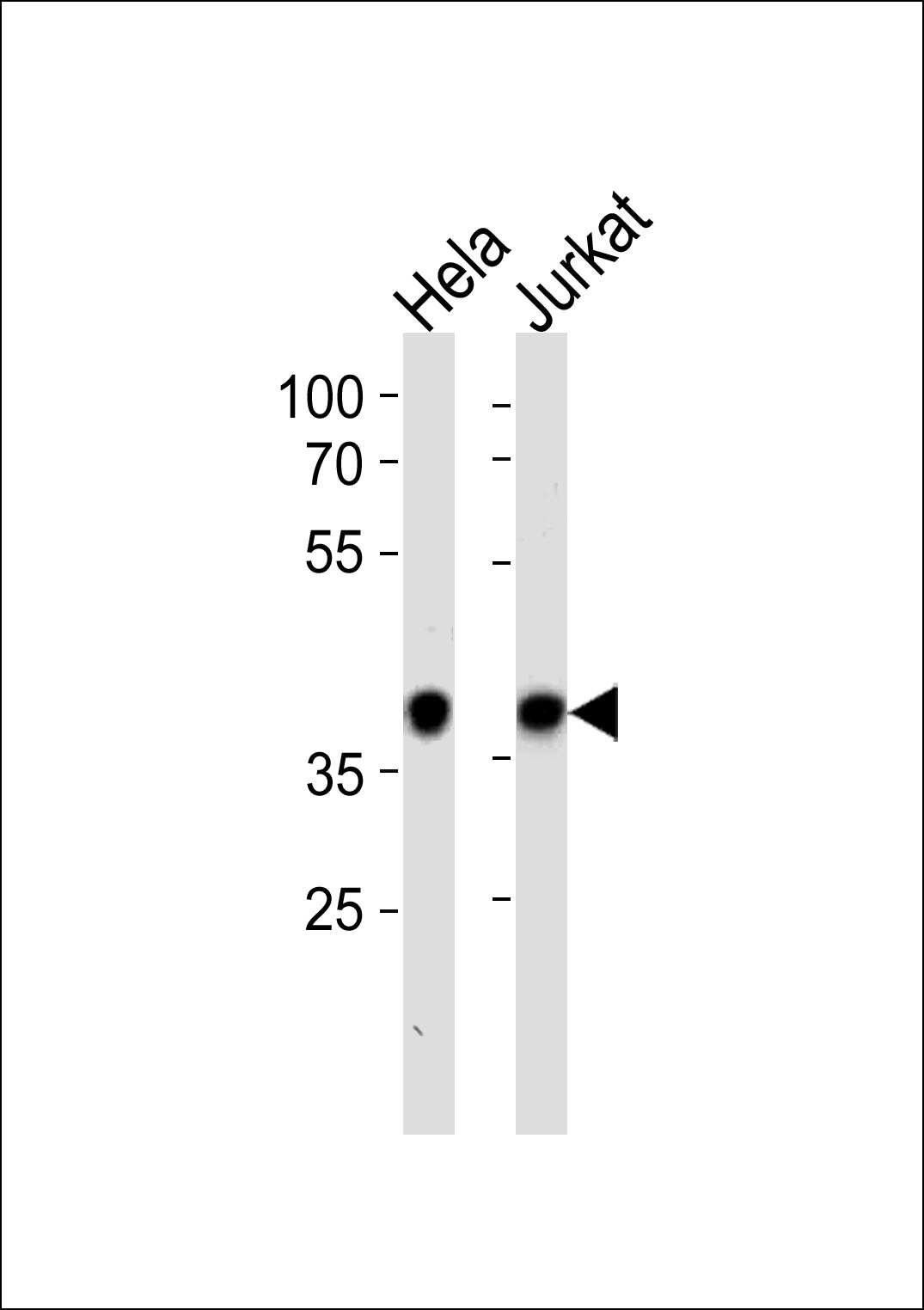
NPM1 Antibody (C-term) (C) western blot analysis in Hela,Jurkat cell line lysates (35ug/lane).This demonstrates the NPM1 antibody detected the NPM1 protein (arrow).| Product Name | Nucleophosmin antibody, C-term |
|---|---|
| Antibody Type | Primary Antibodies |
| Product description | Rabbit polyclonal to NPM1 (C-term)This gene encodes a phosphoprotein which moves between the nucleus and the cytoplasm. The gene product is thought to be involved in several processes including regulation of the ARF/p53 pathway. A number of genes are fusion partners have been characterized, in particular the anaplastic lymphoma kinase gene on chromosome 2. Mutations in this gene are associated with acute myeloid leukemia. More than a dozen pseudogenes of this gene have been identified. Alternative splicing results in multiple transcript variants. |
| Immunogen | KLH conjugated synthetic peptide between 205~234 amino acids from the C-terminal region of human NPM1. |
| Clonality | Polyclonal |
|---|---|
| Isotype | IgG |
| Host Species | Rabbit |
| Tested Applications | FACSICC/IFIHC-PWB |
| ELISA: Use at 1:1,000: |
|
| Species Reactivity | Human |
| Concentration | 0.2mg/ml |
| Gene Synonyms | Nucleolar phosphoprotein B23 Nucleophosmin Numatrin Nucleolar protein NO38 NPM NPM1 |
|---|---|
| Target | NPM1 |
| Molecular Weight(MW) | 32.575 kDa (note) |
| Cellular Localization | Generally nucleolar, but is translocated to the nucleoplasm in case of serum starvation or treatment with anticancer drugs. Has been found in the cytoplasm in patients with primary acute myelogenous leukemia (AML), but not with secondary AML. Can shuttle |
Application
NPM1 Antibody (C-term) flow cytometric analysis of Hela cells (right histogram) compared to a negative control cell (left histogram).FITC-conjugated goat-anti-rabbit secondary antibodies were used for the analysis.
Application
Fluorescent image of A549 cell stained with NPM1 Antibody (C-term).A549 cells were fixed with 4% PFA (20 min), permeabilized with Triton X-100 (0.1%, 10 min), then incubated with NPM1 primary antibody (1:25, 1 h at 37℃). For secondary antibody, Alexa Fluor® 488 conjugated donkey anti-rabbit antibody (green) was used (1:400, 50 min at 37℃).Cytoplasmic actin was counterstained with Alexa Fluor® 555 (red) conjugated Phalloidin (7units/ml, 1 h at 37℃).NPM1 immunoreactivity is localized to Nucleus and Nucleolus significantly.
Application
NPM1 Antibody (C-term) immunohistochemistry analysis in formalin fixed and paraffin embedded human colon carcinoma followed by peroxidase conjugation of the secondary antibody and DAB staining.This data demonstrates the use of NPM1 Antibody (C-term) for immunohistochemistry. Clinical relevance has not been evaluated.
Application
NPM1 Antibody (C-term) (C) western blot analysis in Hela,Jurkat cell line lysates (35ug/lane).This demonstrates the NPM1 antibody detected the NPM1 protein (arrow).| Application Notes | ELISA: Use at 1:1,000: |
|---|
| Form | Liquid |
|---|---|
| Storage Instructions | Store at at 2-8°C for up to 6 months. For long term storage, aliquot and store at -20 °C. Avoid repeated freeze/thaw cycles. |
| Storage Buffer | PBS with 0.09% (W/V) sodium azide |


