Application
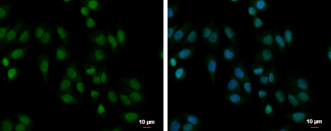
PABPN1 antibody detects PABPN1 protein at cytoplasm and nucleus by immunofluorescent analysis. Sample: A375 cells were fixed in 4% paraformaldehyde at RT for 15 min. Green: PABPN1 protein stained by PABPN1 antibody diluted at 1:500. Blue: Hoechst 33342 staining. Scale bar = 10 μm.Application
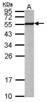
Sample (50 ug of whole cell lysate) A: mouse brain 12% SDS PAGE diluted at 1:1000Application
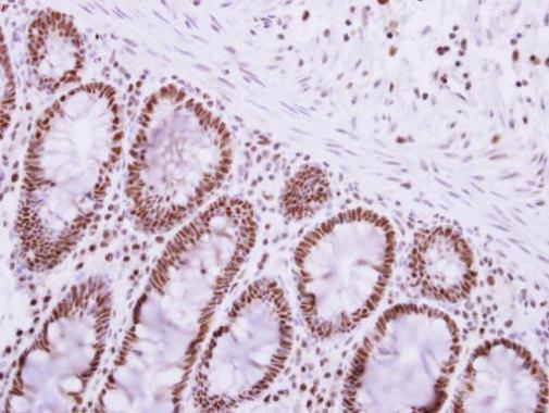
Immunohistochemical analysis of paraffin-embedded human colon carcinoma, using PABPN1 antibody at 1:250 dilution.Application
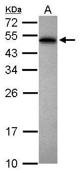
Sample (30 ug of whole cell lysate) A: Jurkat 12% SDS PAGE diluted at 1:1000| Product Name | PABPN1 antibody |
|---|---|
| Antibody Type | Primary Antibodies |
| Product description | Rabbit Polyclonal antibody to PABPN1 (poly(A) binding protein, nuclear 1)This gene encodes an abundant nuclear protein that binds with high affinity to nascent poly(A) tails. The protein is required for progressive and efficient polymerization of poly(A) tails on the 3' ends of eukaryotic genes and controls the size of the poly(A) tail to about 250 nt. At steady-state, this protein is localized in the nucleus whereas a different poly(A) binding protein is localized in the cytoplasm. An expansion of the trinucleotide (GCG) repeat from normal 6 to 8-13 at the 5' end of the coding region of this gene leads to autosomal dominant oculopharyngeal muscular dystrophy (OPMD) disease. Multiple splice variants have been described but their full-length nature is not known. One splice variant includes introns 1 and 6 but no protein is formed. [provided by RefSeq] |
| Immunogen | Recombinant protein encompassing a sequence within the center region of human PABPN1. The exact sequence is proprietary. |
| Clonality | Polyclonal |
|---|---|
| Isotype | IgG |
| Host Species | Rabbit |
| Tested Applications | ICC/IF,IHC-F,IHC-P,WB |
| *Optimal dilutions/concentrations should be determined by the researcher. | |
| Species Reactivity | Human,Mouse |
| Predicted Reactivity | Rat, Zebrafish, Xenopus laevis, Dog, Pig, BovineRat (99%), Zebrafish (83%), Xenopus laevis (88%), Dog (100%), Pig (100%), Bovine (100%) |
| Concentration | 1mg/ml |
| Purification | Affinity purified |
| Gene Synonyms | OPMD antibody, PAB2 antibody, PABII antibody, PABP-2 antibody, PABP2 antibody, PABPN1 antibody, poly(A) binding protein II antibody, polyadenylate-binding protein 2 antibody, poly(A) binding protein 2 antibody, "poly(A) binding protein, nuclear 1 antibody" |
|---|---|
| Molecular Weight(MW) | 33 kDa (note) |
| Cellular Localization | Nucleus (By similarity) , Cytoplasm (By similarity) |
Application
PABPN1 antibody detects PABPN1 protein at cytoplasm and nucleus by immunofluorescent analysis. Sample: A375 cells were fixed in 4% paraformaldehyde at RT for 15 min. Green: PABPN1 protein stained by PABPN1 antibody diluted at 1:500. Blue: Hoechst 33342 staining. Scale bar = 10 μm.
Application
Sample (50 ug of whole cell lysate) A: mouse brain 12% SDS PAGE diluted at 1:1000
Application
Immunohistochemical analysis of paraffin-embedded human colon carcinoma, using PABPN1 antibody at 1:250 dilution.
Application
Sample (30 ug of whole cell lysate) A: Jurkat 12% SDS PAGE diluted at 1:1000| Positive Control | Jurkat , Raji , mouse brain |
|---|---|
| Application Notes | *Optimal dilutions/concentrations should be determined by the researcher. |
| Form | Liquid |
|---|---|
| Storage Instructions | Keep as concentrated solution. Aliquot and store at -20ºC or below. Avoid multiple freeze-thaw cycles. |
| Storage Buffer | 1XPBS, 1% BSA, 20% Glycerol (pH7). 0.01% Thimerosal was added as a preservative. |


