Application
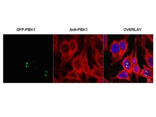
Immunofluorescence microscopy of HeLa cells transfected with GFP-PBK1. In the overlay, specific antibody staining is shown to co-localize with recombinant protein. Cells were fixed with methanol prior to staining.Application
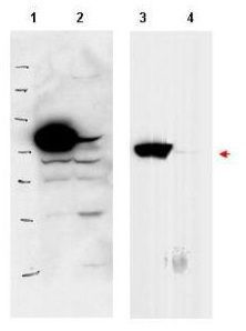
Western blot using GeneTex's affinity purified anti-PBK1 antibody shows detection of over-expressed PBK1 in lysates from HeLa cells transfected with Flag-PBK1. Lanes 1 and 3 contain lysate from Flag-PBK1 transfected HeLa cells. Lanes 2 and 4 contain lysate from cells transfected with vector. Lanes 1 and 2 were blotted with anti-Flag antibody. Lanes 3 and 4 were probed with a 1:500 dilution of anti-PBK1. The band at 75 kDa, indicated by the arrowhead, corresponds to PBK1.| Product Name | Pbk1 antibody |
|---|---|
| Antibody Type | Primary Antibodies |
| Product description | Rabbit Polyclonal antibody to Pbk1PBK1 protein (also known as Ribosomal L1 domain-containing protein 1, cellular senescence-inhibited gene protein, and CATX-11) was isolated from highly invasive first trimester trophoblast cells and has been proposed to regulate their naturally occurring invasive behavior (Huch et al., 1998). PBK1 was also found to be over-expressed in non-small-cell lung cancer (NSCLC) cells (Petroziello et al., 2004).A recent study suggests that PBK1 may up-regulate the urokinase-type plasminogen activator (uPA) gene, which plays an important role in cellular matrix degradation and |
| Immunogen | This affinity purified antibody was prepared from whole rabbit serum produced by repeated immunizations with a synthetic peptide corresponding to an internal sequence of human PBK1. |
| Clonality | Polyclonal |
|---|---|
| Isotype | IgG |
| Host Species | Rabbit |
| Tested Applications | ELISAICC/IFIPWB |
| *Optimal dilutions/concentrations should be determined by the researcher.
: |
|
| Species Reactivity | Human |
| Concentration | 1mg/ml |
| Purification | Affinity purified |
| Gene Synonyms | CSIG L12 PBK1 UTP30 RSL1D1 |
|---|---|
| Target | Pbk1 |
Application
Immunofluorescence microscopy of HeLa cells transfected with GFP-PBK1. In the overlay, specific antibody staining is shown to co-localize with recombinant protein. Cells were fixed with methanol prior to staining.
Application
Western blot using GeneTex's affinity purified anti-PBK1 antibody shows detection of over-expressed PBK1 in lysates from HeLa cells transfected with Flag-PBK1. Lanes 1 and 3 contain lysate from Flag-PBK1 transfected HeLa cells. Lanes 2 and 4 contain lysate from cells transfected with vector. Lanes 1 and 2 were blotted with anti-Flag antibody. Lanes 3 and 4 were probed with a 1:500 dilution of anti-PBK1. The band at 75 kDa, indicated by the arrowhead, corresponds to PBK1.| Application Notes | *Optimal dilutions/concentrations should be determined by the researcher.
: |
|---|
| Form | Liquid |
|---|---|
| Storage Instructions | Store vial at -20° C prior to opening. Aliquot contents and freeze at -20° C or below for extended storage. Avoid cycles of freezing and thawing. |
| Storage Buffer | 0.02 M Potassium Phosphate, 0.15 M Sodium Chloride, pH 7.2 and 0.01% (w/v) Sodium Azide |


