Application
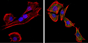
Immunofluorescent analysis of PC2 (green) showing staining in the cytoplasm of C2C12 cells. Formalin-fixed cells were permeabilized with 0.1% Triton X-100 in TBS for 5-10 minutes and blocked with 3% BSA-PBS for 30 minutes at room temperature. Cells were probed with a PC2 polyclonal antibody in 3% BSA-PBS at a dilution of 1:100 and incubated overnight at 4 ºC in a humidified chamber. Cells were washed with PBST and incubated with a DyLight-conjugated secondary antibody in PBS at room temperature in the dark. F-actin (red) was stained with a flourescent red phalloidin and nuclei (blue) were stained with Hoechst or DAPI. Images were taken at a magnification of 60x.Application
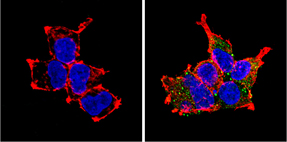
Immunofluorescent analysis of PC2 (green) showing staining in the cytoplasm of HEK293 cells. Formalin-fixed cells were permeabilized with 0.1% Triton X-100 in TBS for 5-10 minutes and blocked with 3% BSA-PBS for 30 minutes at room temperature. Cells were probed with a PC2 polyclonal antibody in 3% BSA-PBS at a dilution of 1:100 and incubated overnight at 4 ºC in a humidified chamber. Cells were washed with PBST and incubated with a DyLight-conjugated secondary antibody in PBS at room temperature in the dark. F-actin (red) was stained with a flourescent red phalloidin and nuclei (blue) were stained with Hoechst or DAPI. Images were taken at a magnification of 60x.| Product Name | PC2 antibody |
|---|---|
| Antibody Type | Primary Antibodies |
| Product description | Rabbit Polyclonal antibody to PC2The subtilisin-like prohormone convertase (PC) family is a group of cellular enzymes that cleave most prohormones and neuropeptide precursors. Numerous other cellular proteins, some viral proteins, and bacterial toxins that are transported by the constitutive secretory pathway are also targeted for maturation by PCs. PC family members share structural similarities, which include a heterogeneous ~10 kDa amino-terminal proregion, a highly conserved ~55 kDa subtilisin-like catalytic domain, and carboxyl-terminal domain that is heterogeneous in length and sequence. These enzymes become catalytically active following proregion cleavage within the appropriate cellular compartment. The subcellular localization of PC family members varies. Immunolocalization studies show that PC1 is found in the perinuclear region as well as the trans-Golgi network, whereas PC2 can be found in the trans-Golgi network as well as diffusely distributed in the peripheral cytoplasm. |
| Immunogen | Synthetic peptide corresponding to residues E(622) L D E A V E R S L Q S I L R K N(638) of mouse PC2. |
| Clonality | Polyclonal |
|---|---|
| Host Species | Rabbit |
| Tested Applications | ICC/IFWB |
| ICC/IF: 1:20 - 1:200: |
|
| Species Reactivity | HumanMouse |
| Concentration | 1mg/ml (Please refer to the vial label for the specific concentration) |
| Purification | Affinity purified |
| Gene Synonyms | Pcsk2 AI839700 SPC2 Phpp-2 6330411F23Rik Phpp2 Nec2 Nec-2 Prohormone Convertase2 PC2 Prohormone Convertase 2 18549 Nec 2 Phpp 2 |
|---|---|
| Target | PC2 |
Application
Immunofluorescent analysis of PC2 (green) showing staining in the cytoplasm of C2C12 cells. Formalin-fixed cells were permeabilized with 0.1% Triton X-100 in TBS for 5-10 minutes and blocked with 3% BSA-PBS for 30 minutes at room temperature. Cells were probed with a PC2 polyclonal antibody in 3% BSA-PBS at a dilution of 1:100 and incubated overnight at 4 ºC in a humidified chamber. Cells were washed with PBST and incubated with a DyLight-conjugated secondary antibody in PBS at room temperature in the dark. F-actin (red) was stained with a flourescent red phalloidin and nuclei (blue) were stained with Hoechst or DAPI. Images were taken at a magnification of 60x.
Application
Immunofluorescent analysis of PC2 (green) showing staining in the cytoplasm of HEK293 cells. Formalin-fixed cells were permeabilized with 0.1% Triton X-100 in TBS for 5-10 minutes and blocked with 3% BSA-PBS for 30 minutes at room temperature. Cells were probed with a PC2 polyclonal antibody in 3% BSA-PBS at a dilution of 1:100 and incubated overnight at 4 ºC in a humidified chamber. Cells were washed with PBST and incubated with a DyLight-conjugated secondary antibody in PBS at room temperature in the dark. F-actin (red) was stained with a flourescent red phalloidin and nuclei (blue) were stained with Hoechst or DAPI. Images were taken at a magnification of 60x.| Application Notes | ICC/IF: 1:20 - 1:200: |
|---|
| Form | Liquid |
|---|---|
| Storage Instructions | Keep as concentrated solution. Aliquot and store at -20ºC or below. Avoid multiple freeze-thaw cycles. |
| Storage Buffer | PBS containing 1 mg/ml BSA and 0.05% sodium azide. |


