Application
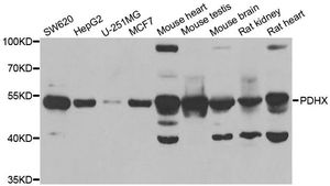
WB analysis of extracts from various cells using PDHX antibody.Application
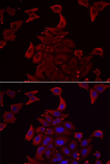
ICC/IF analysis of U2OS cells using PDHX antibody (red) and DAPI (blue).| Product Name | PDHX antibody |
|---|---|
| Antibody Type | Primary Antibodies |
| Product description | Rabbit Polyclonal antibody to PDHXThe pyruvate dehydrogenase (PDH) complex is located in the mitochondrial matrix and catalyzes the conversion of pyruvate to acetyl coenzyme A. The PDH complex thereby links glycolysis to Krebs cycle. The PDH complex contains three catalytic subunits, E1, E2, and E3, two regulatory subunits, E1 kinase and E1 phosphatase, and a non-catalytic subunit, E3 binding protein (E3BP). This gene encodes the E3 binding protein subunit; also known as component X of the pyruvate dehydrogenase complex. This protein tethers E3 dimers to the E2 core of the PDH complex. Defects in this gene are a cause of pyruvate dehydrogenase deficiency which results in neurological dysfunction and lactic acidosis in infancy and early childhood. This protein is also a minor antigen for antimitochondrial antibodies. These autoantibodies are present in nearly 95% of patients with the autoimmune liver disease primary biliary cirrhosis (PBC). In PBC, activated T lymphocytes attack and destroy epithelial cells in the bile |
| Immunogen | Recombinant protein of human PDHX |
| Clonality | Polyclonal |
|---|---|
| Isotype | IgG |
| Host Species | Rabbit |
| Tested Applications | ICC/IFIHC-PWB |
| WB: 1:500 - 1:2000. IHC: 1:50 - 1:200: |
|
| Species Reactivity | Human |
| Concentration | 1mg/ml |
| Purification | Affinity purified |
| Gene Synonyms | DLDBP E3BP proX OPDX PDX1 pyruvate dehydrogenase complex component X |
|---|---|
| Target | PDHX |
| Molecular Weight(MW) | 54 kDa (note) |
Application
WB analysis of extracts from various cells using PDHX antibody.
Application
ICC/IF analysis of U2OS cells using PDHX antibody (red) and DAPI (blue).| Application Notes | WB: 1:500 - 1:2000. IHC: 1:50 - 1:200: |
|---|
| Form | Liquid |
|---|---|
| Storage Instructions | Store at -20°C (regular) or -80°C (long term). Avoid freeze / thaw cycles. |
| Storage Buffer | PBS with 0.02% sodium azide, 50% glycerol, pH7.3. |


