Application
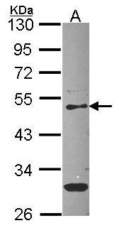
Sample (30 ug of whole cell lysate) A: Hep G2 10% SDS PAGE PEDF antibody diluted at 1:1000Application
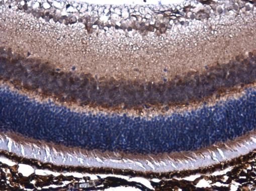
PEDF antibody [N1C2] detects PEDF protein at cytoplasm in mouse eye by immunohistochemical analysis. Sample: Paraffin-embedded mouse eye. PEDF antibody [N1C2] diluted at 1:400.WB
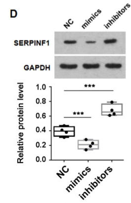
(D) Western blot results showing transfection of Agomir (mimics) and Antagomir (inhibitors) of miR-186-5p suppressed and de-repressed, respectively, SERPINF1 protein expression in HT-22 cells.WB
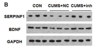
(B) Western blot showing SERPINF1 and brain-derived neurotrophic factor (BDNF) protein expression in hippocampus of CUMS mice following tail intravenous injection of Antagomir inhibitors (n = 3 mice per group).WB
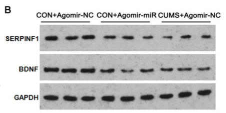
(B) Western blot showing SERPINF1 and BDNF protein expression in hippocampus of wild-type mice following tail intravenous injection of Agomir-miR-186-5p.IHC
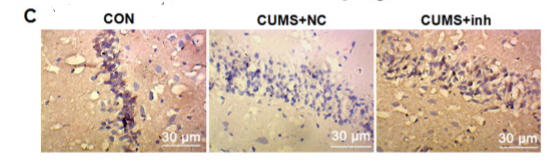
(C) Representative IHC results and statistics showing specifically decreased SERPINF1 protein in hippocampus of CUMS mice, which was significantly restored upon systemic injection of Antagomir miR-186-5p.IHC
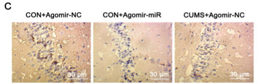
(C) Representative IHC results and statistics showing that the hippocampal SERPINF1 protein expression in wild-type mice was repressed to a level comparable to that of in CUMS mice.| Product Name | PEDF antibody |
|---|---|
| Antibody Type | Primary Antibodies |
| Product description | Rabbit polyclonal antibody to PEDF (serpin peptidase inhibitor, clade F (alpha-2 antiplasmin, pigment epithelium derived factor), member 1)The PEDF gene is a member of the serpin gene family. Serpins are a group of serine protease inhibitors, some of which have also been reported to exhibit neurotrophic activity.[supplied by OMIM] |
| Immunogen | Recombinant protein encompassing a sequence within the center region of human PEDF. The exact sequence is proprietary. |
| Clonality | Polyclonal |
|---|---|
| Isotype | IgG |
| Host Species | Rabbit |
| Tested Applications | IHC-PWB |
| *Optimal dilutions/concentrations should be determined by the researcher.
: |
|
| Species Reactivity | HumanMouse |
| Predicted Reactivity | Rat, Dog, Pig, Sheep, Bovine, Guinea pigRat (83%), Dog (87%), Pig (87%), Sheep (86%), Bovine (86%), Guinea pig (83%) |
| Concentration | 1mg/ |
| Purification | Affinity purified |
| Gene Synonyms | EPC-1 antibody PEDF antibody SERPINF1 antibody pigment epithelium-derived factor antibody cell proliferation-inducing gene 35 protein antibody "serine (or cysteine) proteinase inhibitor clade F (alpha-2 antiplasmin pigment epithelium derived factor) |
|---|---|
| Target | PEDF |
| Molecular Weight(MW) | 46 kDa (note) (3) |
| Cellular Localization | Secreted , Melanosome |
Application
Sample (30 ug of whole cell lysate) A: Hep G2 10% SDS PAGE PEDF antibody diluted at 1:1000
Application
PEDF antibody [N1C2] detects PEDF protein at cytoplasm in mouse eye by immunohistochemical analysis. Sample: Paraffin-embedded mouse eye. PEDF antibody [N1C2] diluted at 1:400.
WB
(D) Western blot results showing transfection of Agomir (mimics) and Antagomir (inhibitors) of miR-186-5p suppressed and de-repressed, respectively, SERPINF1 protein expression in HT-22 cells.
WB
(B) Western blot showing SERPINF1 and brain-derived neurotrophic factor (BDNF) protein expression in hippocampus of CUMS mice following tail intravenous injection of Antagomir inhibitors (n = 3 mice per group).
WB
(B) Western blot showing SERPINF1 and BDNF protein expression in hippocampus of wild-type mice following tail intravenous injection of Agomir-miR-186-5p.
IHC
(C) Representative IHC results and statistics showing specifically decreased SERPINF1 protein in hippocampus of CUMS mice, which was significantly restored upon systemic injection of Antagomir miR-186-5p.
IHC
(C) Representative IHC results and statistics showing that the hippocampal SERPINF1 protein expression in wild-type mice was repressed to a level comparable to that of in CUMS mice.| Positive Control | A431 , H1299 , HeLa , HepG2 |
|---|---|
| Application Notes | *Optimal dilutions/concentrations should be determined by the researcher.
: |
| Form | Liquid |
|---|---|
| Storage Instructions | Keep as concentrated solution. Aliquot and store at -20ºC or below. Avoid multiple freeze-thaw cycles. |
| Storage Buffer | 0.1M Tris, 0.1M Glycine, 20% Glycerol (pH7). 0.01% Thimerosal was added as a preservative. |


