Application
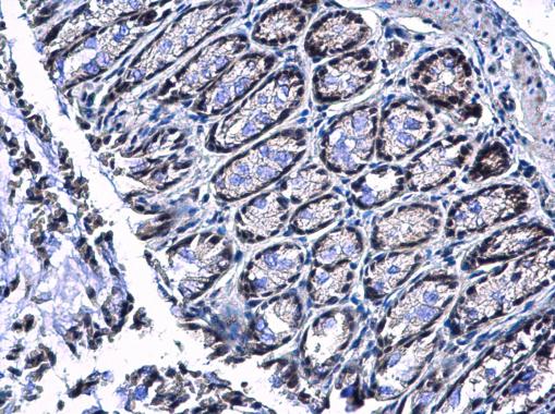
PIASy antibody detects PIASy protein at nucleus in mouse colon by immunohistochemical analysis. Sample: Paraffin-embedded mouse colon. PIASy antibody diluted at 1:500.Application
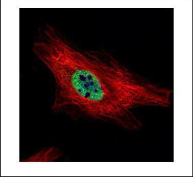
Confocal immunofluorescence analysis (Olympus FV10i) of paraformaldehyde-fixed HeLa, using PIASy antibody (Green) at 1:500 dilution. Alpha-tubulin filaments were labeled with (Red) at 1:2000.Application
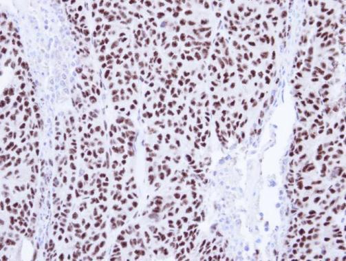
Immunohistochemical analysis of paraffin-embedded DLD1 Xenograft, using PIAS4 antibody at 1:100 dilution.Application
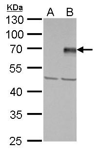
PIASy antibody detects PIASy protein by western blot analysis. A. 30 µg 293T whole cell lysate/extract B. 30 µg whole cell lysate/extract of human PIAS4-transfected 293T cells 10% SDS-PAGE PIASy antibody dilution: 1:5000 The HRP-conjugated anti-rabbit IgG antibody was used to detect the primary antibody.| Product Name | PIASy antibody |
|---|---|
| Antibody Type | Primary Antibodies |
| Product description | Rabbit Polyclonal antibody to PIASy (protein inhibitor of activated STAT, 4)Functions as an E3-type small ubiquitin-like modifier (SUMO) ligase, stabilizing the interaction between UBE2I and the substrate, and as a SUMO-tethering factor. Plays a crucial role as a transcriptional coregulation in various cellular pathways, including the STAT pathway, the p53 pathway, the Wnt pathway and the steroid hormone signaling pathway. Involved in gene silencing. Promotes PARK7 sumoylation. In Wnt signaling, represses LEF1 and enhances TCF4 transcriptional activities through promoting their sumoylations. |
| Immunogen | Carrier-protein conjugated synthetic peptide encompassing a sequence within the C-terminus region of human PIASy. The exact sequence is proprietary. |
| Clonality | Polyclonal |
|---|---|
| Isotype | IgG |
| Host Species | Rabbit |
| Tested Applications | ICC/IFIHC-PWB |
| *Optimal dilutions/concentrations should be determined by the researcher. | |
| Species Reactivity | HumanMouse |
| Predicted Reactivity | Chimpanzee, BovineChimpanzee (100%), Bovine (100%) |
| Concentration | 1mg/ml (Please refer to the vial label for the specific concentration) |
| Purification | Affinity purified |
| Gene Synonyms | FLJ12419 antibody, MGC35296 antibody, PIASY antibody, Piasg antibody, ZMIZ6 antibody, PIAS4 antibody, protein inhibitor of activated STAT protein PIASy antibody, PIAS-gamma antibody, "zinc finger, MIZ-type containing 6 antibody", protein inhibitor of activ |
|---|---|
| Molecular Weight(MW) | 57 kDa (note) |
| Cellular Localization | Nucleus |
Application
PIASy antibody detects PIASy protein at nucleus in mouse colon by immunohistochemical analysis. Sample: Paraffin-embedded mouse colon. PIASy antibody diluted at 1:500.
Application
Confocal immunofluorescence analysis (Olympus FV10i) of paraformaldehyde-fixed HeLa, using PIASy antibody (Green) at 1:500 dilution. Alpha-tubulin filaments were labeled with (Red) at 1:2000.
Application
Immunohistochemical analysis of paraffin-embedded DLD1 Xenograft, using PIAS4 antibody at 1:100 dilution.
Application
PIASy antibody detects PIASy protein by western blot analysis. A. 30 µg 293T whole cell lysate/extract B. 30 µg whole cell lysate/extract of human PIAS4-transfected 293T cells 10% SDS-PAGE PIASy antibody dilution: 1:5000 The HRP-conjugated anti-rabbit IgG antibody was used to detect the primary antibody.| Application Notes | *Optimal dilutions/concentrations should be determined by the researcher. |
|---|
| Form | Liquid |
|---|---|
| Storage Instructions | Keep as concentrated solution. Aliquot and store at -20ºC or below. Avoid multiple freeze-thaw cycles. |
| Storage Buffer | 0.1M Tris, 0.1M Glycine, 10% Glycerol (pH7). 0.01% Thimerosal was added as a preservative. |


