Application
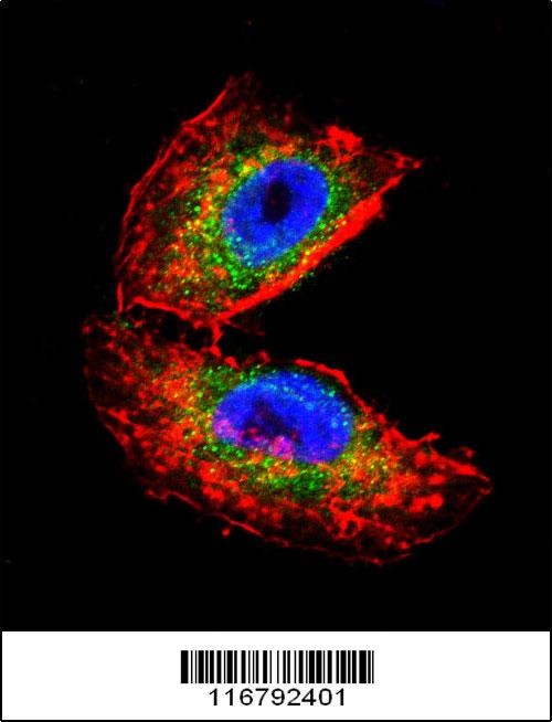
Confocal immunofluorescent analysis of ACHE Antibody with NCI-H460 cell followed by Alexa Fluor 488-conjugated goat anti-rabbit lgG (green).Actin filaments have been labeled with Alexa Fluor 555 phalloidin (red).DAPI was used to stain the cell nuclear (blue).Application
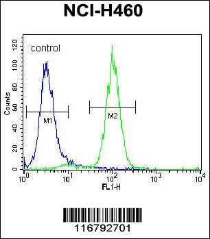
Flow cytometric analysis of NCI-H460 cells (right histogram) compared to a negative control cell (left histogram).FITC-conjugated goat-anti-rabbit secondary antibodies were used for the analysis.Application
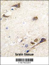
Formalin-fixed and paraffin-embedded human brain tissue reacted with ACHE antibody (N-term), which was peroxidase-conjugated to the secondary antibody, followed by DAB staining.Application
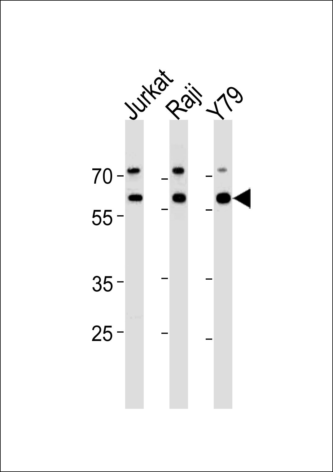
Western blot analysis in Jurkat,Raji,Y79 cell line lysates (35ug/lane).| Product Name | ACHE Antibody |
|---|---|
| Antibody Type | Primary Antibodies |
| Antigen Alias | Acetylcholinesterase, AChE, ACHE |
| Product description | Acetylcholinesterase hydrolyzes the neurotransmitter, acetylcholine at neuromuscular junctions and brain cholinergic synapses, and thus terminates signal transmission. The Protein is also found on the red blood cell membranes, where it constitutes the Yt blood group antigen. Acetylcholinesterase exists in multiple molecular forms which possess similar catalytic properties, but differ in their oligomeric assembly and mode of cell attachment to the cell surface. The major form of acetylcholinesterase found in brain, muscle and other tissues is the hydrophilic species, which forms disulfide-linked oligomers with collagenous, or lipid-containing structural subunits.1) Liang,D., FEBS J. 276 (1), 94-108 (2009) |
| Immunogen | This ACHE antibody is generated from rabbits immunized with a KLH conjugated synthetic peptide between 147-175 amino acids from the N-terminal region of human ACHE. |
| Clonality | Polyclonal |
|---|---|
| Isotype | Ig |
| Host Species | Rabbit |
| Tested Applications | FACSIFIHC-PWB |
| For WB starting dilution is: 1:1000 For IHC-P starting dilution is: 1:10~50 For FACS starting dilution is: 1:10~50 For IF starting dilution is: 1:10~50: |
|
| Species Reactivity | Human |
| Concentration | 1mg/ml |
| Gene Symbol | ACHE |
|---|---|
| Alternative Names | Acetylcholinesterase AChE ACHE |
| Molecular Weight(MW) | 68 kDa |
| Sequence Similarities | Predicted species reactivity based on immunogen sequence: Guinea Pig |
Application
Confocal immunofluorescent analysis of ACHE Antibody with NCI-H460 cell followed by Alexa Fluor 488-conjugated goat anti-rabbit lgG (green).Actin filaments have been labeled with Alexa Fluor 555 phalloidin (red).DAPI was used to stain the cell nuclear (blue).
Application
Flow cytometric analysis of NCI-H460 cells (right histogram) compared to a negative control cell (left histogram).FITC-conjugated goat-anti-rabbit secondary antibodies were used for the analysis.
Application
Formalin-fixed and paraffin-embedded human brain tissue reacted with ACHE antibody (N-term), which was peroxidase-conjugated to the secondary antibody, followed by DAB staining.
Application
Western blot analysis in Jurkat,Raji,Y79 cell line lysates (35ug/lane).| Application Notes | For WB starting dilution is: 1:1000 For IHC-P starting dilution is: 1:10~50 For FACS starting dilution is: 1:10~50 For IF starting dilution is: 1:10~50: |
|---|
| Form | Liquid |
|---|---|
| Storage Instructions | Store at 4˚C for three months and -20˚C, stable for up to one year. As with all antibodies care should be taken to avoid repeated freeze thaw cycles. Antibodies should not be exposed to prolonged high temperatures. |
| Storage Buffer | Supplied in PBS with 0.09% (W/V) sodium azide. |


