Application
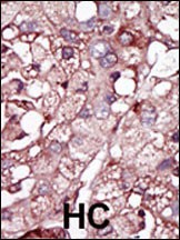
Formalin-fixed and paraffin-embedded human cancer tissue reacted with the primary antibody, which was peroxidase-conjugated to the secondary antibody, followed by DAB staining. BC = breast carcinoma; HC = hepatocarcinoma.Application
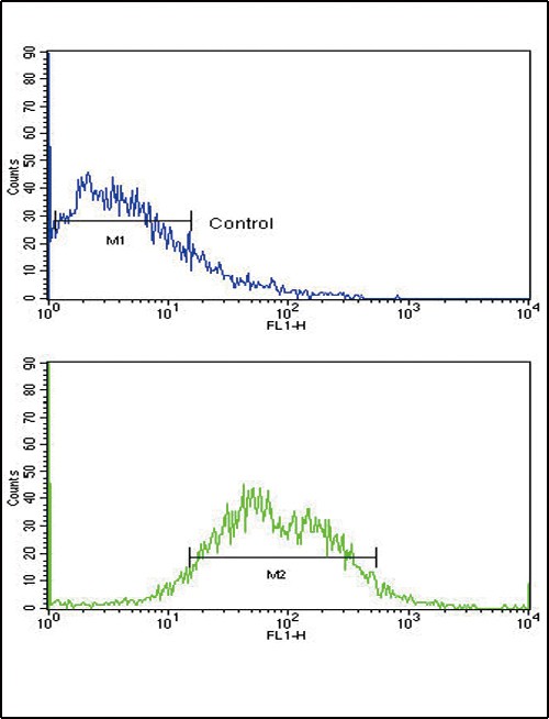
Flow cytometric analysis of HepG2 cells using ACVRL1 Antibody (bottom histogram) compared to a negative control cell (top histogram). FITC-conjugated goat-anti-rabbit secondary antibodies were used for the analysis.Application
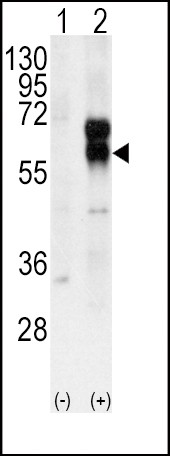
Western blot analysis of ACVRL1 using rabbit polyclonal ACVRL1 Antibody using 293 cell lysates (2 ug/lane) either nontransfected (Lane 1) or transiently transfected with the ACVRL1 gene (Lane 2).Application
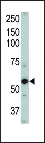
Western blot of ACVRL1 Pab . TOP LEFT: Mouse heart tissue lysate.| Product Name | ACVRL1 Antibody |
|---|---|
| Antibody Type | Primary Antibodies |
| Antigen Alias | Serine/threonine-protein kinase receptor R3, SKR3, Activin receptor-like kinase 1, ALK-1, TGF-B superfamily receptor type I, TSR-I, ACVRL1, ACVRLK1, ALK1 |
| Product description | ACVRL1 is a type I cell-surface receptor for the TGF-beta superfamily of ligands. It shares with other type I receptors a high degree of similarity in serine-threonine kinase subdomains, a glycine- and serine-rich region (called the GS domain) preceding the kinase domain, and a short C-terminal tail. This protein, sometimes termed ALK1, shares similar domain structures with other closely related ALK or activin receptor-like kinase proteins that form a subfamily of receptor serine/threonine kinases. Mutations in this gene are associated with hemorrhagic telangiectasia type 2, also known as Rendu-Osler-Weber syndrome 2.1) Strausberg, R.L., et al., Proc. Natl. Acad. Sci. U.S.A. 99(26):16899-16903 (2002). |
| Immunogen | This ACVRL1 antibody is generated from rabbits immunized with a KLH conjugated synthetic peptide between 38-68 amino acids from the N-terminal region of human ACVRL1. |
| Clonality | Polyclonal |
|---|---|
| Isotype | Ig |
| Host Species | Rabbit |
| Tested Applications | FACSIHC-PWB |
| For WB starting dilution is: 1:1000 For FACS starting dilution is: 1:10~50 For IHC-P starting dilution is: 1:50~100: |
|
| Species Reactivity | HumanMouse |
| Concentration | 1mg/ml |
| Gene Symbol | ACVRL1 |
|---|---|
| Alternative Names | Serine/threonine-protein kinase receptor R3 SKR3 Activin receptor-like kinase 1 ALK-1 TGF-B superfamily receptor type I TSR-I ACVRL1 ACVRLK1 ALK1 |
| Molecular Weight(MW) | 56 kDa |
Application
Formalin-fixed and paraffin-embedded human cancer tissue reacted with the primary antibody, which was peroxidase-conjugated to the secondary antibody, followed by DAB staining. BC = breast carcinoma; HC = hepatocarcinoma.
Application
Flow cytometric analysis of HepG2 cells using ACVRL1 Antibody (bottom histogram) compared to a negative control cell (top histogram). FITC-conjugated goat-anti-rabbit secondary antibodies were used for the analysis.
Application
Western blot analysis of ACVRL1 using rabbit polyclonal ACVRL1 Antibody using 293 cell lysates (2 ug/lane) either nontransfected (Lane 1) or transiently transfected with the ACVRL1 gene (Lane 2).
Application
Western blot of ACVRL1 Pab . TOP LEFT: Mouse heart tissue lysate.| Application Notes | For WB starting dilution is: 1:1000 For FACS starting dilution is: 1:10~50 For IHC-P starting dilution is: 1:50~100: |
|---|
| Form | Liquid |
|---|---|
| Storage Instructions | Store at 4˚C for three months and -20˚C, stable for up to one year. As with all antibodies care should be taken to avoid repeated freeze thaw cycles. Antibodies should not be exposed to prolonged high temperatures. |
| Storage Buffer | Supplied in PBS with 0.09% (W/V) sodium azide. |


