Application
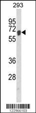
Western blot analysis of AIFM1 Antibody in 293 cell line lysates (35ug/lane)Application
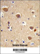
Formalin-fixed and paraffin-embedded human brain tissue reacted with AIFM1 Antibody (N-term), which was peroxidase-conjugated to the secondary antibody, followed by DAB staining.Application
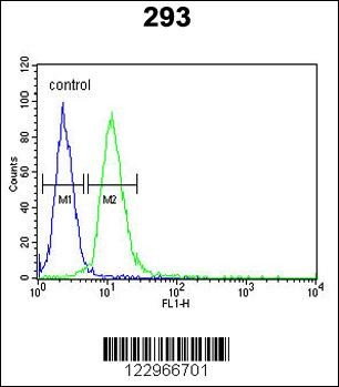
Flow cytometric analysis of 293 cells (right histogram) compared to a negative control cell (left histogram).FITC-conjugated goat-anti-rabbit secondary antibodies were used for the analysis.Application
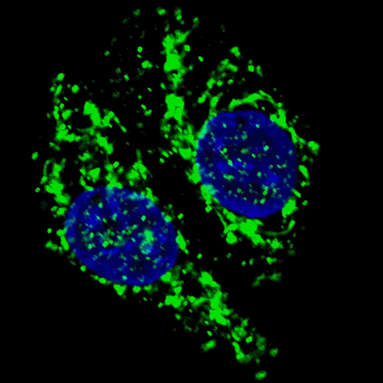
Fluorescent confocal image of U251 cells stained with AIFM1 antibody. U251 cells were treated with Chloroquine (50 uM,16h), then fixed with 4% PFA (20 min), permeabilized with Triton X-100 (0.2%, 30 min). Cells were then incubated with AP8910a AIFM1 primary antibody (1:200, 2 h at room temperature). For secondary antibody, Alexa Fluor 488 conjugated donkey anti-rabbit antibody (green) was used (1:Application
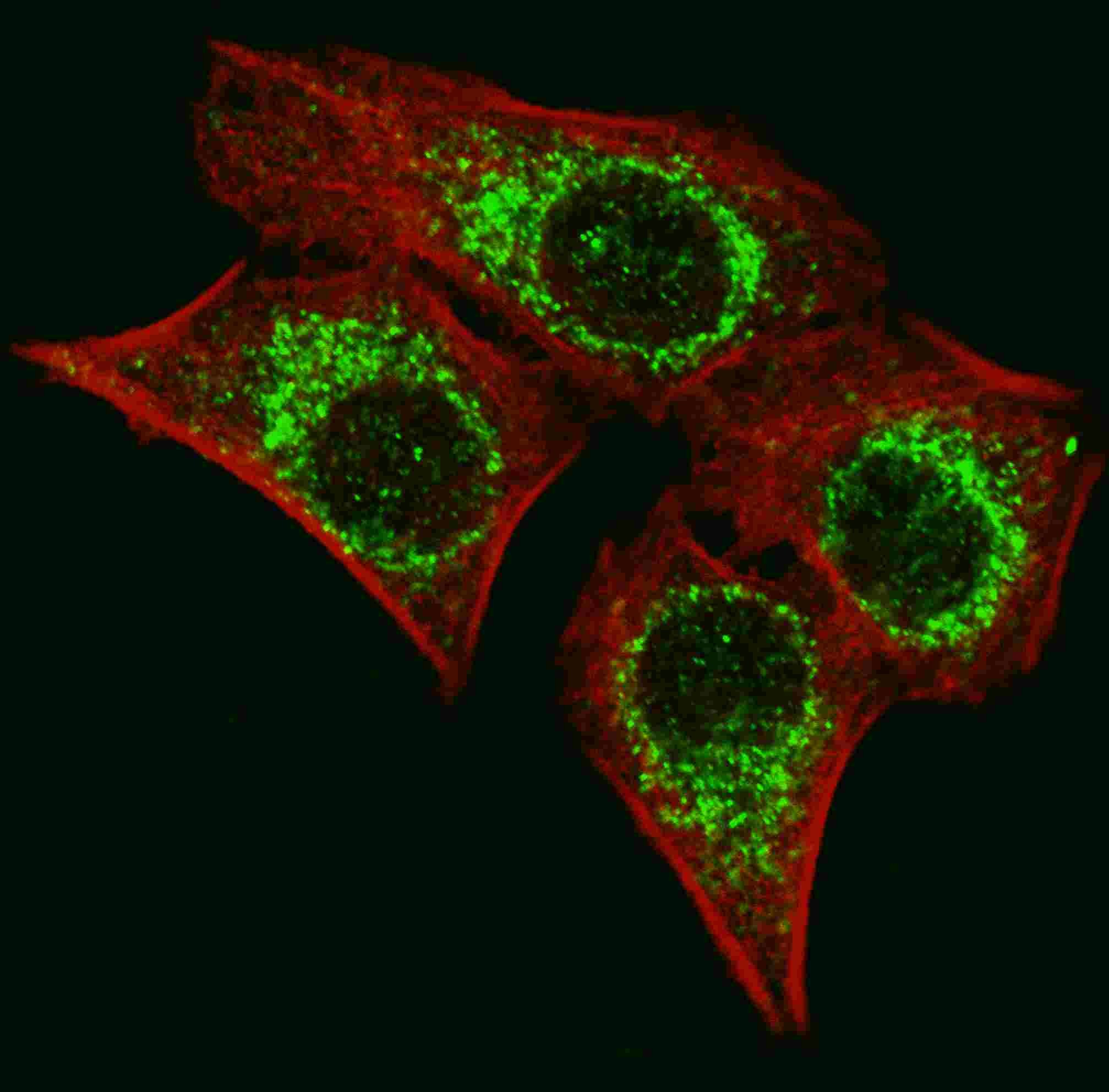
Fluorescent image of U251 cells stained with AIFM1 antibody. U251 cells were fixed with 4% PFA (20 min), permeabilized with Triton X-100 (0.2%, 30 min). Cells were then incubated with AIFM1 primary antibody (1:200, 2 h at room temperature). For secondary antibody, Alexa Fluor 488 conjugated donkey anti-rabbit antibody (green) was used (1:1000, 1h). Cytoplasmic actin was counterstained with Alexa F| Product Name | AIFM1 Antibody |
|---|---|
| Antibody Type | Primary Antibodies |
| Antigen Alias | Apoptosis-inducing factor 1, mitochondrial, 111-, Programmed cell death protein 8, AIFM1, AIF, PDCD8 |
| Product description | AIFM1 is a flavoprotein essential for nuclear disassembly in apoptotic cells that is found in the mitochondrial intermembrane space in healthy cells. Induction of apoptosis results in the translocation of this protein to the nucleus where it effects chromosome condensation and fragmentation. In addition, this protein induces mitochondria to release the apoptogenic proteins cytochrome c and caspase-9.1) References for protein: |
| Immunogen | This AIFM1 antibody is generated from rabbits immunized with a KLH conjugated synthetic peptide between 70-98 amino acids from the N-terminal region of human AIFM1. |
| Clonality | Polyclonal |
|---|---|
| Isotype | Ig |
| Host Species | Rabbit |
| Tested Applications | FACSIFIHC-PWB |
| For IF starting dilution is: 1:200 For WB starting dilution is: 1:1000 For IHC-P starting dilution is: 1:50~100 For FACS starting dilution is: 1:10~50: |
|
| Species Reactivity | Human |
| Concentration | 1mg/ml |
| Purification | Affinity purified |
| Gene Symbol | AIFM1 |
|---|---|
| Alternative Names | Apoptosis-inducing factor 1 mitochondrial 111- Programmed cell death protein 8 AIFM1 AIF PDCD8 |
| Molecular Weight(MW) | 67 kDa |
Application
Western blot analysis of AIFM1 Antibody in 293 cell line lysates (35ug/lane)
Application
Formalin-fixed and paraffin-embedded human brain tissue reacted with AIFM1 Antibody (N-term), which was peroxidase-conjugated to the secondary antibody, followed by DAB staining.
Application
Flow cytometric analysis of 293 cells (right histogram) compared to a negative control cell (left histogram).FITC-conjugated goat-anti-rabbit secondary antibodies were used for the analysis.
Application
Fluorescent confocal image of U251 cells stained with AIFM1 antibody. U251 cells were treated with Chloroquine (50 uM,16h), then fixed with 4% PFA (20 min), permeabilized with Triton X-100 (0.2%, 30 min). Cells were then incubated with AP8910a AIFM1 primary antibody (1:200, 2 h at room temperature). For secondary antibody, Alexa Fluor 488 conjugated donkey anti-rabbit antibody (green) was used (1:
Application
Fluorescent image of U251 cells stained with AIFM1 antibody. U251 cells were fixed with 4% PFA (20 min), permeabilized with Triton X-100 (0.2%, 30 min). Cells were then incubated with AIFM1 primary antibody (1:200, 2 h at room temperature). For secondary antibody, Alexa Fluor 488 conjugated donkey anti-rabbit antibody (green) was used (1:1000, 1h). Cytoplasmic actin was counterstained with Alexa F| Application Notes | For IF starting dilution is: 1:200 For WB starting dilution is: 1:1000 For IHC-P starting dilution is: 1:50~100 For FACS starting dilution is: 1:10~50: |
|---|
| Form | Liquid |
|---|---|
| Storage Instructions | Store at 4˚C for three months and -20˚C, stable for up to one year. As with all antibodies care should be taken to avoid repeated freeze thaw cycles. Antibodies should not be exposed to prolonged high temperatures. |
| Storage Buffer | Supplied in PBS with 0.09% (W/V) sodium azide. |


 Data sheet for OM270528