Application
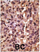
Formalin-fixed and paraffin-embedded human cancer tissue reacted with the primary antibody, which was peroxidase-conjugated to the secondary antibody, followed by DAB staining. BC = breast carcinoma; HC = hepatocarcinoma.Application
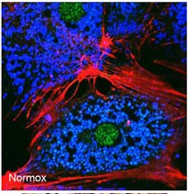
Freshly isolated mouse hepatocytes plated on coverslips (2 x10⁵ cells/22-mm glass coverslip) were cultured under normoxic conditions for 6 hr. The cells were then fixed in 2% paraformaldehyde in PBS for 1 hr, and processed for confocal immunofluorescence (red: F-actin, blue: ATP-synthase, green: BNIP3). Fluorescence labeling of BNIP3 accomplished with anti-BNIP3 antibody. Data courtesy of Ruben ZaApplication
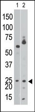
Antibody is used in Western blot to detect NIP3 BH3 in Ramos cell lysate (lane 1) and in mouse brain tissue lysate (lane 2).Application
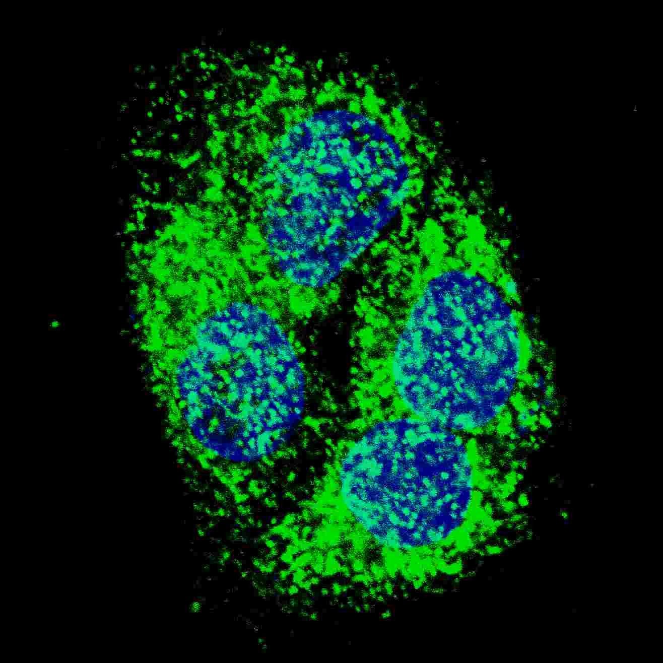
Fluorescent confocal image of HepG2 cells stained with BNIP3 (BH3 Domain Specific) antibody. HepG2 cells were fixed with 4% PFA (20 min), permeabilized with Triton X-100 (0.2%, 30 min). Cells were then incubated with BNIP3 (BH3 Domain Specific) primary antibody (1:500, 2 h at room temperature). For secondary antibody, Alexa Fluor 488 conjugated donkey anti-rabbit antibody (green) was used (1:1000,| Product Name | BNIP3 Antibody |
|---|---|
| Antibody Type | Primary Antibodies |
| Product description | NIP3 is a member of the BCL2/adenovirus E1B 19 kd-interacting protein (BNIP) family. It interacts with the E1B 19 kDa protein which is responsible for the protection of virally-induced cell death, as well as E1B 19 kDa-like sequences of BCL2, also an apoptotic protector. NIP3 contains a BH3 domain and a transmembrane domain, which have been associated with pro-apoptotic function. The dimeric mitochondrial protein is known to induce apoptosis, even in the presence of BCL2.1) References for protein: |
| Immunogen | This BNIP3 antibody is generated from rabbits immunized with a KLH conjugated synthetic peptide between 152-187 amino acids from human BNIP3. |
| Clonality | Polyclonal |
|---|---|
| Isotype | Ig |
| Host Species | Rabbit |
| Tested Applications | IFIHC-PWB |
| For IF starting dilution is: 1:500 For WB starting dilution is: 1:1000 For IHC-P starting dilution is: 1:50~100 | |
| Species Reactivity | HumanMouse |
| Concentration | 1mg/ml |
| Purification | Unpurified |
| Gene Symbol | BNIP3 |
|---|---|
| Alternative Names | BCL2/adenovirus E1B 19 kDa protein-interacting protein 3 BNIP3 NIP3 |
| Molecular Weight(MW) | 22 kDa |
Application
Formalin-fixed and paraffin-embedded human cancer tissue reacted with the primary antibody, which was peroxidase-conjugated to the secondary antibody, followed by DAB staining. BC = breast carcinoma; HC = hepatocarcinoma.
Application
Freshly isolated mouse hepatocytes plated on coverslips (2 x10⁵ cells/22-mm glass coverslip) were cultured under normoxic conditions for 6 hr. The cells were then fixed in 2% paraformaldehyde in PBS for 1 hr, and processed for confocal immunofluorescence (red: F-actin, blue: ATP-synthase, green: BNIP3). Fluorescence labeling of BNIP3 accomplished with anti-BNIP3 antibody. Data courtesy of Ruben Za
Application
Antibody is used in Western blot to detect NIP3 BH3 in Ramos cell lysate (lane 1) and in mouse brain tissue lysate (lane 2).
Application
Fluorescent confocal image of HepG2 cells stained with BNIP3 (BH3 Domain Specific) antibody. HepG2 cells were fixed with 4% PFA (20 min), permeabilized with Triton X-100 (0.2%, 30 min). Cells were then incubated with BNIP3 (BH3 Domain Specific) primary antibody (1:500, 2 h at room temperature). For secondary antibody, Alexa Fluor 488 conjugated donkey anti-rabbit antibody (green) was used (1:1000,| Application Notes | For IF starting dilution is: 1:500 For WB starting dilution is: 1:1000 For IHC-P starting dilution is: 1:50~100 |
|---|
| Form | Liquid |
|---|---|
| Storage Instructions | Store at 4˚C for three months and -20˚C, stable for up to one year. As with all antibodies care should be taken to avoid repeated freeze thaw cycles. Antibodies should not be exposed to prolonged high temperatures. |
| Storage Buffer | Supplied in PBS with 0.09% (W/V) sodium azide. |


