Application
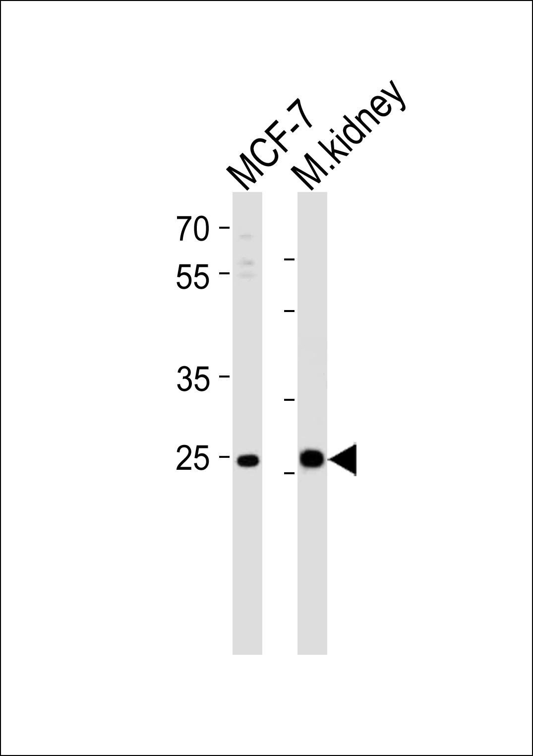
Western blot analysis of lysates from MCF-7 cell line and mouse kidney tissue lysate (from left to right), using CA2 Antibody at 1:1000 at each lane.Application
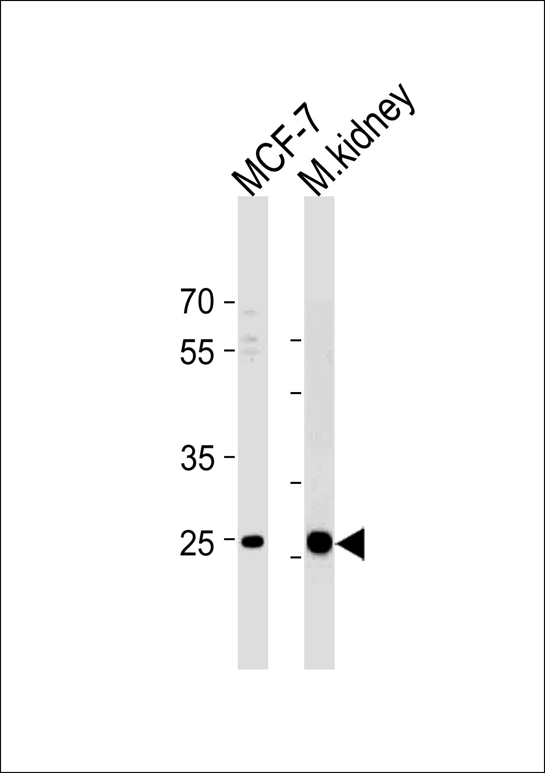
Western blot analysis in MCF-7 cell line and mouse kidney tissue lysates (35ug/lane).Application
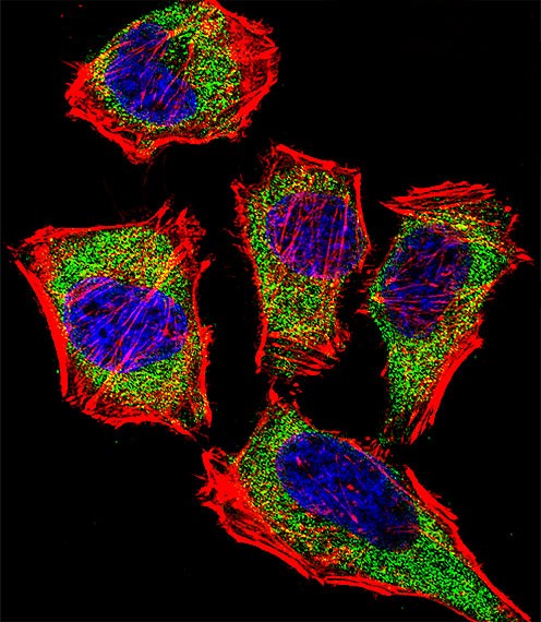
Fluorescent confocal image of Hela cell stained with CA2 Antibody (N-term).Hela cells were fixed with 4% PFA (20 min), permeabilized with Triton X-100 (0.1%, 10 min), then incubated with CA2 primary antibody (1:25). For secondary antibody, Alexa Fluor 488 conjugated donkey anti-rabbit antibody (green) was used (1:400).Cytoplasmic actin was counterstained with Alexa Fluor 555 (red) conjugated Phall| Product Name | CA2 Antibody |
|---|---|
| Antibody Type | Primary Antibodies |
| Product description | CA2 is one of several (at least 7) isozymes of carbonic anhydrase. Carbonic anhydrase catalyzes reversible hydration of carbon dioxide. Defects in this enzyme are associated with osteopetrosis and renal tubular acidosis.1) Sultana, R., et al. Antioxid. Redox Signal. 12(3):327-336(2010) |
| Immunogen | This CA2 antibody is generated from rabbits immunized with a KLH conjugated synthetic peptide between 54-83 amino acids from the N-terminal region of human CA2. |
| Clonality | Polyclonal |
|---|---|
| Isotype | Ig |
| Host Species | Rabbit |
| Tested Applications | IFWB |
| For WB starting dilution is: 1:1000 For IF starting dilution is: 1:10~50: |
|
| Species Reactivity | HumanMouse |
| Concentration | 1mg/ml |
| Gene Symbol | CA2 |
|---|---|
| Alternative Names | Carbonic anhydrase 2 Carbonate dehydratase II Carbonic anhydrase C CAC Carbonic anhydrase II CA-II CA2 |
| Molecular Weight(MW) | 29 kDa |
Application
Western blot analysis of lysates from MCF-7 cell line and mouse kidney tissue lysate (from left to right), using CA2 Antibody at 1:1000 at each lane.
Application
Western blot analysis in MCF-7 cell line and mouse kidney tissue lysates (35ug/lane).
Application
Fluorescent confocal image of Hela cell stained with CA2 Antibody (N-term).Hela cells were fixed with 4% PFA (20 min), permeabilized with Triton X-100 (0.1%, 10 min), then incubated with CA2 primary antibody (1:25). For secondary antibody, Alexa Fluor 488 conjugated donkey anti-rabbit antibody (green) was used (1:400).Cytoplasmic actin was counterstained with Alexa Fluor 555 (red) conjugated Phall| Application Notes | For WB starting dilution is: 1:1000 For IF starting dilution is: 1:10~50: |
|---|
| Form | Liquid |
|---|---|
| Storage Instructions | Store at 4˚C for three months and -20˚C, stable for up to one year. As with all antibodies care should be taken to avoid repeated freeze thaw cycles. Antibodies should not be exposed to prolonged high temperatures. |
| Storage Buffer | Supplied in PBS with 0.09% (W/V) sodium azide. |


