Application
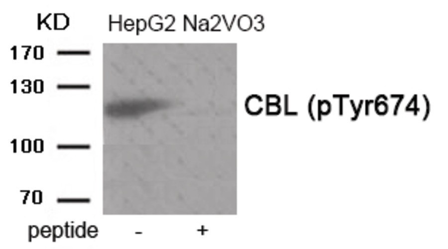
Western blot analysis of extracts from HepG2 cells treated with Na2VO3 using Phospho-CBL (Tyr674) Antibody. The lane on the right is treated with the antigen-specific peptide.| Product Name | CBL (phospho Tyr674) Antibody |
|---|---|
| Antibody Type | Primary Antibodies |
| Antigen Alias | C-CBL, CBL2, Proto-oncogene c-Cbl, RNF55, |
| Product description | Participates in signal transduction in hematopoietic cells. Adapter protein that functions as a negative regulator of many signaling pathways that start from receptors at the cell surface. Acts as an E3 ubiquitin-protein ligase, which accepts ubiquitin from specific E2 ubiquitin-conjugating enzymes, and then transfers it to substrates promoting their degradation by the proteasome. Recognizes activated receptor tyrosine kinases, including PDGFA, EGF and CSF1, and terminates signaling.1) Fernandes MS, et al. (2010) J Biol Chem 285, 32596-605 |
| Immunogen | CBL (phospho Tyr674) antibody was raised against a peptide sequence around phosphorylation site of Tyrosine674 (A-I-Y (p)-S-L) derived from Human CBL. |
| Modification | p-Tyr674 |
| Clonality | Polyclonal |
|---|---|
| Isotype | IgG |
| Host Species | Rabbit |
| Tested Applications | WB |
| Western Blot: 1:500~1:1000: |
|
| Species Reactivity | HumanMouseRat |
| Concentration | 1mg/ml |
| Purification | Affinity purified |
| Gene Symbol | CBL |
|---|---|
| Alternative Names | C-CBL CBL2 Proto-oncogene c-Cbl RNF55 |
| Molecular Weight(MW) | 120 kDa |
| Tissue Specificity | The antibody detects endogenous level of CBL only when phosphorylated at tyrosine 674. |
| Entrez Gene | 867 |
|---|---|
| Protein Accession | NP_005179.2 |
Application
Western blot analysis of extracts from HepG2 cells treated with Na2VO3 using Phospho-CBL (Tyr674) Antibody. The lane on the right is treated with the antigen-specific peptide.| Application Notes | Western Blot: 1:500~1:1000: |
|---|
| Form | Liquid |
|---|---|
| Storage Instructions | Store antibody at -20˚C for up to one year. |
| Storage Buffer | Antibody supplied in phosphate buffered saline (without Mg2+ and Ca2+), pH 7.4, 150mM NaCl, 0.02% sodium azide and 50% glycerol. |


