Application
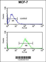
Flow cytometric analysis of MCF-7 cells (bottom histogram) compared to a negative control cell (top histogram).FITC-conjugated goat-anti-rabbit secondary antibodies were used for the analysis.Application
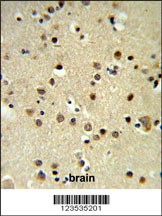
CBX1 Antibody IHC analysis in formalin fixed and paraffin embedded brain tissue followed by peroxidase conjugation of the secondary antibody and DAB staining.Application
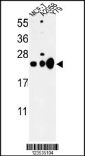
Western blot analysis of CBX1 Antibody in MCF-7, A2058, Y79 cell line lysates (35ug/lane)| Product Name | CBX1 Antibody |
|---|---|
| Antibody Type | Primary Antibodies |
| Antigen Alias | Chromobox protein homolog 1, HP1Hsbeta, Heterochromatin protein 1 homolog beta, HP1 beta, Heterochromatin protein p25, M31, Modifier 1 protein, p25beta, CBX1, CBX |
| Product description | CBX1 is a highly conserved nonhistone protein, which is a member of the heterochromatin protein family . The protein is enriched in the heterochromatin and associated with centromeres. The protein has a single N-terminal chromodomain which can bind to histone proteins via methylated lysine residues, and a C-terminal chromo shadow-domain (CSD) which is responsible for the homodimerization and interaction with a number of chromatin-associated nonhistone proteins. The protein may play an important role in the epigenetic control of chromatin structure and gene expression.1) Aagaard,L., et.al., EMBO J. 18 (7), 1923-1938 (1999) |
| Immunogen | This CBX1 antibody is generated from rabbits immunized with a KLH conjugated synthetic peptide between 128-155 amino acids from the C-terminal region of human CBX1. |
| Clonality | Polyclonal |
|---|---|
| Isotype | Ig |
| Host Species | Rabbit |
| Tested Applications | FACSIHC-PWB |
| For WB starting dilution is: 1:1000 For IHC-P starting dilution is: 1:50~100 For FACS starting dilution is: 1:10~50: |
|
| Species Reactivity | Human |
| Concentration | 1mg/ml |
| Purification | Affinity purified |
| Gene Symbol | CBX1 |
|---|---|
| Alternative Names | Chromobox protein homolog 1 HP1Hsbeta Heterochromatin protein 1 homolog beta HP1 beta Heterochromatin protein p25 M31 Modifier 1 protein p25beta CBX1 CBX |
| Molecular Weight(MW) | 21 kDa |
| Sequence Similarities | Predicted species reactivity based on immunogen sequence: Mouse |
Application
Flow cytometric analysis of MCF-7 cells (bottom histogram) compared to a negative control cell (top histogram).FITC-conjugated goat-anti-rabbit secondary antibodies were used for the analysis.
Application
CBX1 Antibody IHC analysis in formalin fixed and paraffin embedded brain tissue followed by peroxidase conjugation of the secondary antibody and DAB staining.
Application
Western blot analysis of CBX1 Antibody in MCF-7, A2058, Y79 cell line lysates (35ug/lane)| Application Notes | For WB starting dilution is: 1:1000 For IHC-P starting dilution is: 1:50~100 For FACS starting dilution is: 1:10~50: |
|---|
| Form | Liquid |
|---|---|
| Storage Instructions | Store at 4˚C for three months and -20˚C, stable for up to one year. As with all antibodies care should be taken to avoid repeated freeze thaw cycles. Antibodies should not be exposed to prolonged high temperatures. |
| Storage Buffer | Supplied in PBS with 0.09% (W/V) sodium azide. |


