Application
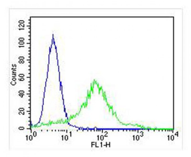
Overlay histogram showing Hela cells stained with Antibody (green line). The cells were fixed with 2% paraformaldehyde (10 min) and then permeabilized with 90% methanol for 10 min. The cells were then icubated in 2% bovine serum albumin to block non-specific protein-protein interactions followed by the antibody (1:25 dilution) for 60 min at 37ºC. The secondary antibody used was Goat-Anti-Rabbit IgApplication
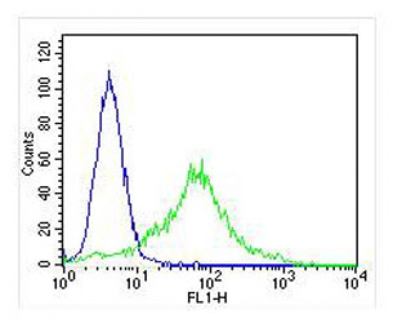
Overlay histogram showing Hela cells stained with Antibody (green line). The cells were fixed with 2% paraformaldehyde (10 min) and then permeabilized with 90% methanol for 10 min. The cells were then icubated in 2% bovine serum albumin to block non-specific protein-protein interactions followed by the antibody (1:25 dilution) for 60 min at 37ºC. The secondary antibody used was Goat-Anti-Rabbit IgApplication
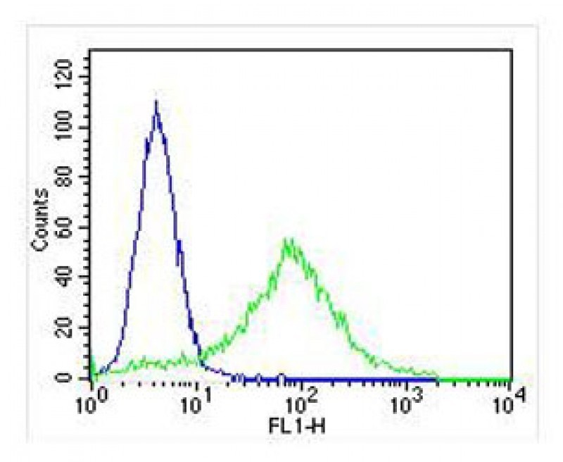
Overlay histogram showing Hela cells stained with Antibody (green line). The cells were fixed with 2% paraformaldehyde (10 min) and then permeabilized with 90% methanol for 10 min. The cells were then icubated in 2% bovine serum albumin to block non-specific protein-protein interactions followed by the antibody (1:25 dilution) for 60 min at 37ºC. The secondary antibody used was Goat-Anti-Rabbit IgApplication
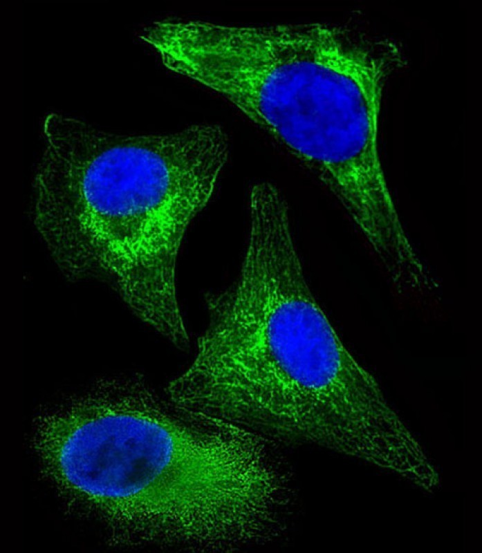
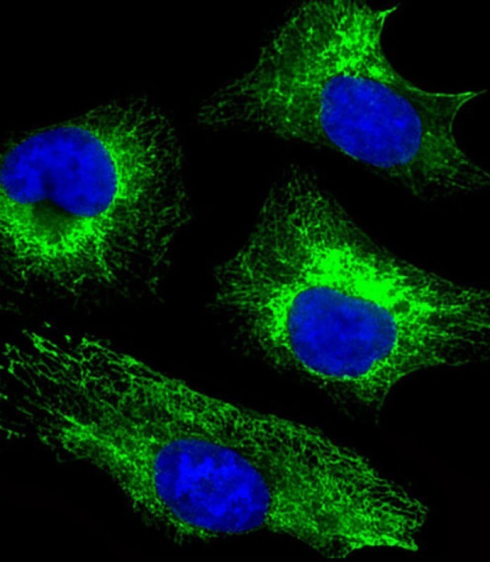
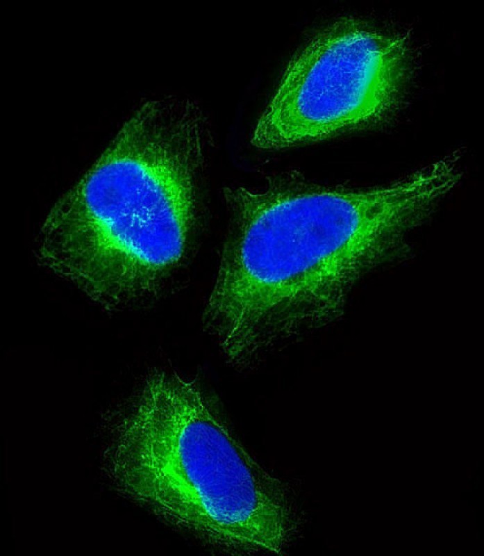
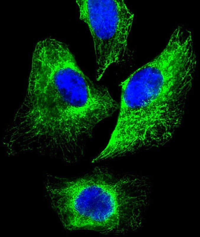
Immunofluorescent analysis of 4% paraformaldehyde-fixed, 0. 1% Triton X-100 permeabilized HeLa (human cervical epithelial adenocarcinoma cell line) cells labeling Pdx1 with antibody at 1/25 dilution, followed by Dylight 488-conjugated goat anti-rabbit IgG (NK179883) secondary antibody at 1/200 dilution (green). Immunofluorescence image showing cytoskeleton staining on HeLa cell line. The nuclear cApplication
Immunofluorescent analysis of 4% paraformaldehyde-fixed, 0. 1% Triton X-100 permeabilized HeLa (human cervical epithelial adenocarcinoma cell line) cells labeling Pdx1 with antibody at 1/25 dilution, followed by Dylight 488-conjugated goat anti-rabbit IgG (NK179883) secondary antibody at 1/200 dilution (green). Immunofluorescence image showing cytoskeleton staining on HeLa cell line. The nuclear cApplication
Immunofluorescent analysis of 4% paraformaldehyde-fixed, 0. 1% Triton X-100 permeabilized HeLa (human cervical epithelial adenocarcinoma cell line) cells labeling Pdx1 with antibody at 1/25 dilution, followed by Dylight 488-conjugated goat anti-rabbit IgG (NK179883) secondary antibody at 1/200 dilution (green). Immunofluorescence image showing cytoskeleton staining on HeLa cell line. The nuclear cApplication
Immunofluorescent analysis of 4% paraformaldehyde-fixed, 0. 1% Triton X-100 permeabilized HeLa (human cervical epithelial adenocarcinoma cell line) cells labeling Pdx1 with antibody at 1/25 dilution, followed by Dylight 488-conjugated goat anti-rabbit IgG (NK179883) secondary antibody at 1/200 dilution (green). Immunofluorescence image showing cytoskeleton staining on HeLa cell line. The nuclear c| Product Name | CYK18 Antibody |
|---|---|
| Antibody Type | Primary Antibodies |
| Product description | KRT18 is the type I intermediate filament chain keratin 18. Keratin 18, together with its filament partner keratin 8, are perhaps the most commonly found members of the intermediate filament family. They are expressed in single layer epithelial tissues of the body. Mutations in its gene have been linked to cryptogenic cirrhosis.1) Zhang,Q., Clin. Cancer Res. 15 (10), 3557-3567 (2009) |
| Immunogen | This CYK18 antibody is generated from rabbits immunized with a KLH conjugated synthetic peptide between 401-430 amino acids from the C-terminal region of human CYK18. |
| Clonality | Polyclonal |
|---|---|
| Isotype | Ig |
| Host Species | Rabbit |
| Tested Applications | FACSIFIHC-PWB |
| For IF starting dilution is: 1:25 For FACS starting dilution is: 1:25 | |
| Species Reactivity | HumanMouse |
| Concentration | 1mg/ml |
| Purification | Unpurified |
| Gene Symbol | KRT18 |
|---|---|
| Alternative Names | Keratin type I cytoskeletal 18 Cell proliferation-inducing gene 46 protein Cytokeratin-18 CK-18 Keratin-18 K18 KRT18 CYK18 |
| Molecular Weight(MW) | 48 kDa |
Application
Overlay histogram showing Hela cells stained with Antibody (green line). The cells were fixed with 2% paraformaldehyde (10 min) and then permeabilized with 90% methanol for 10 min. The cells were then icubated in 2% bovine serum albumin to block non-specific protein-protein interactions followed by the antibody (1:25 dilution) for 60 min at 37ºC. The secondary antibody used was Goat-Anti-Rabbit Ig
Application
Overlay histogram showing Hela cells stained with Antibody (green line). The cells were fixed with 2% paraformaldehyde (10 min) and then permeabilized with 90% methanol for 10 min. The cells were then icubated in 2% bovine serum albumin to block non-specific protein-protein interactions followed by the antibody (1:25 dilution) for 60 min at 37ºC. The secondary antibody used was Goat-Anti-Rabbit Ig
Application
Overlay histogram showing Hela cells stained with Antibody (green line). The cells were fixed with 2% paraformaldehyde (10 min) and then permeabilized with 90% methanol for 10 min. The cells were then icubated in 2% bovine serum albumin to block non-specific protein-protein interactions followed by the antibody (1:25 dilution) for 60 min at 37ºC. The secondary antibody used was Goat-Anti-Rabbit Ig
Application
Immunofluorescent analysis of 4% paraformaldehyde-fixed, 0. 1% Triton X-100 permeabilized HeLa (human cervical epithelial adenocarcinoma cell line) cells labeling Pdx1 with antibody at 1/25 dilution, followed by Dylight 488-conjugated goat anti-rabbit IgG (NK179883) secondary antibody at 1/200 dilution (green). Immunofluorescence image showing cytoskeleton staining on HeLa cell line. The nuclear c
Application
Immunofluorescent analysis of 4% paraformaldehyde-fixed, 0. 1% Triton X-100 permeabilized HeLa (human cervical epithelial adenocarcinoma cell line) cells labeling Pdx1 with antibody at 1/25 dilution, followed by Dylight 488-conjugated goat anti-rabbit IgG (NK179883) secondary antibody at 1/200 dilution (green). Immunofluorescence image showing cytoskeleton staining on HeLa cell line. The nuclear c
Application
Immunofluorescent analysis of 4% paraformaldehyde-fixed, 0. 1% Triton X-100 permeabilized HeLa (human cervical epithelial adenocarcinoma cell line) cells labeling Pdx1 with antibody at 1/25 dilution, followed by Dylight 488-conjugated goat anti-rabbit IgG (NK179883) secondary antibody at 1/200 dilution (green). Immunofluorescence image showing cytoskeleton staining on HeLa cell line. The nuclear c
Application
Immunofluorescent analysis of 4% paraformaldehyde-fixed, 0. 1% Triton X-100 permeabilized HeLa (human cervical epithelial adenocarcinoma cell line) cells labeling Pdx1 with antibody at 1/25 dilution, followed by Dylight 488-conjugated goat anti-rabbit IgG (NK179883) secondary antibody at 1/200 dilution (green). Immunofluorescence image showing cytoskeleton staining on HeLa cell line. The nuclear c| Application Notes | For IF starting dilution is: 1:25 For FACS starting dilution is: 1:25 |
|---|
| Form | Liquid |
|---|---|
| Storage Instructions | Store at 4˚C for three months and -20˚C, stable for up to one year. As with all antibodies care should be taken to avoid repeated freeze thaw cycles. Antibodies should not be exposed to prolonged high temperatures. |
| Storage Buffer | Supplied in PBS with 0.09% (W/V) sodium azide. |


