Application
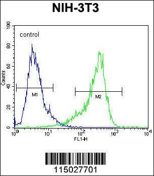
Flow cytometric analysis of NIH-3T3 cells (right histogram) compared to a negative control cell (left histogram).FITC-conjugated goat-anti-rabbit secondary antibodies were used for the analysis.Application
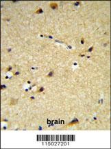
CYP26A1 Antibody IHC analysis in formalin fixed and paraffin embedded brain tissue followed by peroxidase conjugation of the secondary antibody and DAB staining.Application
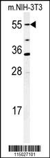
Western blot analysis of CYP26A1 Antibody in mouse NIH-3T3 cell lysates (35ug/lane)| Product Name | CYP26A1 Antibody |
|---|---|
| Antibody Type | Primary Antibodies |
| Product description | CYP26A1 is a member of the cytochrome P450 superfamily of enzymes. The cytochrome P450 proteins are monooxygenases which catalyze many reactions involved in drug metabolism and synthesis of cholesterol, steroids and other lipids.This endoplasmic reticulum protein acts on retinoids, including all-trans-retinoic acid (RA), with both 4-hydroxylation and 18-hydroxylation activities. This enzyme regulates the cellular level of retinoic acid which is involved in regulation of gene expression in both embryonic and adult tissues.1) Quere,R., Blood 109 (10), 4450-4460 (2007) Lee,S.J., Pharmacogenet. Genomics 17 (3), 169-180 (2007) Heise,R., J. Invest. Dermatol. 126 (11), 2473-2480 (2006) |
| Immunogen | This CYP26A1 antibody is generated from rabbits immunized with a KLH conjugated synthetic peptide between 467-497 amino acids from the C-terminal region of human CYP26A1. |
| Clonality | Polyclonal |
|---|---|
| Isotype | Ig |
| Host Species | Rabbit |
| Tested Applications | FACSIHC-PWB |
| For WB starting dilution is: 1:1000 For IHC-P starting dilution is: 1:50~100 For FACS starting dilution is: 1:10~50 | |
| Species Reactivity | HumanMouse |
| Concentration | 1mg/ml |
| Purification | Unpurified |
| Gene Symbol | CYP26A1 |
|---|---|
| Alternative Names | Cytochrome P450 26A1 114-- Cytochrome P450 retinoic acid-inactivating 1 Cytochrome P450RAI hP450RAI Retinoic acid 4-hydroxylase Retinoic acid-metabolizing cytochrome CYP26A1 CYP26 P450RAI1 |
| Molecular Weight(MW) | 56 kDa |
Application
Flow cytometric analysis of NIH-3T3 cells (right histogram) compared to a negative control cell (left histogram).FITC-conjugated goat-anti-rabbit secondary antibodies were used for the analysis.
Application
CYP26A1 Antibody IHC analysis in formalin fixed and paraffin embedded brain tissue followed by peroxidase conjugation of the secondary antibody and DAB staining.
Application
Western blot analysis of CYP26A1 Antibody in mouse NIH-3T3 cell lysates (35ug/lane)| Application Notes | For WB starting dilution is: 1:1000 For IHC-P starting dilution is: 1:50~100 For FACS starting dilution is: 1:10~50 |
|---|
| Form | Liquid |
|---|---|
| Storage Instructions | Store at 4˚C for three months and -20˚C, stable for up to one year. As with all antibodies care should be taken to avoid repeated freeze thaw cycles. Antibodies should not be exposed to prolonged high temperatures. |
| Storage Buffer | Supplied in PBS with 0.09% (W/V) sodium azide. |


