Application
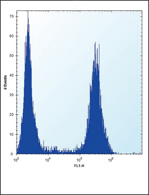
Flow cytometric analysis of U251 cells (right histogram) compared to a negative control cell (left histogram).FITC-conjugated goat-anti-rabbit secondary antibodies were used for the analysis.Application
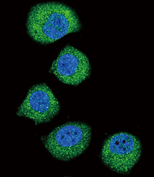
Confocal immunofluorescent analysis of FGFR2 Antibody with U251 cell followed by Alexa Fluor 488-conjugated goat anti-rabbit lgG (green). DAPI was used to stain the cell nuclear (blue).Application
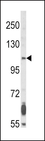
Western blot analysis of FGFR2 Antibody in NCI-H460 cell line lysates (35ug/lane)Application
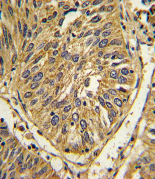
Formalin-fixed and paraffin-embedded human lung carcinoma with FGFR2 Antibody, which was peroxidase-conjugated to the secondary antibody, followed by DAB staining.Application
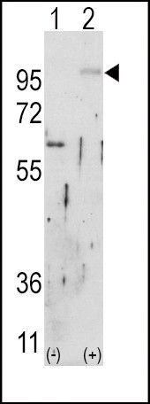
Western blot analysis of FGFR2 using rabbit polyclonal FGFR2 Antibody using 293 cell lysates (2 ug/lane) either nontransfected (Lane 1) or transiently transfected with the FGFR2 gene (Lane 2).| Product Name | FGFR2 Antibody |
|---|---|
| Antibody Type | Primary Antibodies |
| Product description | FGFR2 is a member of the fibroblast growth factor receptor family, where amino acid sequence is highly conserved between members and throughout evolution. FGFR family members differ from one another in their ligand affinities and tissue distribution. A full-length representative protein consists of an extracellular region, composed of three immunoglobulin-like domains, a single hydrophobic membrane-spanning segment and a cytoplasmic tyrosine kinase domain. The extracellular portion of the protein interacts with fibroblast growth factors, setting in motion a cascade of downstream signals, ultimately influencing mitogenesis and differentiation. This particular family member is a high-affinity receptor for acidic, basic and/or keratinocyte growth factor, depending on the isoform. Mutations in FGFR2 gene are associated with Crouzon syndrome, Pfeiffer syndrome, Craniosynostosis, Apert syndrome, Jackson-Weiss syndrome, Beare-Stevenson cutis gyrata syndrome, Saethre-Chotzen syndrome, and syndromic craniosynostosis.1) Park W.-J., Meyers G.A., Li X.Hum. Mol. Genet. 4:1229-1233(1995) |
| Immunogen | This FGFR2 antibody is generated from rabbits immunized with a his tag recombinant protein of human FGFR2. |
| Clonality | Polyclonal |
|---|---|
| Isotype | Ig |
| Host Species | Rabbit |
| Tested Applications | FACSIFIHC-PWB |
| For WB starting dilution is: 1:1000 For IHC-P starting dilution is: 1:10~50 For IF starting dilution is: 1:10~50 For FACS starting dilution is: 1:10~50 | |
| Species Reactivity | Human |
| Concentration | 1mg/ml |
| Purification | Unpurified |
| Gene Symbol | FGFR2 |
|---|---|
| Alternative Names | Fibroblast growth factor receptor 2 FGFR-2 K-sam KGFR Keratinocyte growth factor receptor CD332 FGFR2 BEK KGFR KSAM |
| Molecular Weight(MW) | 92 kDa |
Application
Flow cytometric analysis of U251 cells (right histogram) compared to a negative control cell (left histogram).FITC-conjugated goat-anti-rabbit secondary antibodies were used for the analysis.
Application
Confocal immunofluorescent analysis of FGFR2 Antibody with U251 cell followed by Alexa Fluor 488-conjugated goat anti-rabbit lgG (green). DAPI was used to stain the cell nuclear (blue).
Application
Western blot analysis of FGFR2 Antibody in NCI-H460 cell line lysates (35ug/lane)
Application
Formalin-fixed and paraffin-embedded human lung carcinoma with FGFR2 Antibody, which was peroxidase-conjugated to the secondary antibody, followed by DAB staining.
Application
Western blot analysis of FGFR2 using rabbit polyclonal FGFR2 Antibody using 293 cell lysates (2 ug/lane) either nontransfected (Lane 1) or transiently transfected with the FGFR2 gene (Lane 2).| Application Notes | For WB starting dilution is: 1:1000 For IHC-P starting dilution is: 1:10~50 For IF starting dilution is: 1:10~50 For FACS starting dilution is: 1:10~50 |
|---|
| Form | Liquid |
|---|---|
| Storage Instructions | Store at 4˚C for three months and -20˚C, stable for up to one year. As with all antibodies care should be taken to avoid repeated freeze thaw cycles. Antibodies should not be exposed to prolonged high temperatures. |
| Storage Buffer | Supplied in PBS with 0.09% (W/V) sodium azide. |


 Data sheet for OM278828