Application
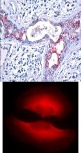
Top Image: Immunohistochemical analysis of paraffin-embedded human breast carcinoma tissue using GSK3β (Phospho-Ser9). Bottom Image: Immunofluorescence staining of methanol-fixed HeLa cells showing cytoplasmic staining using GSK3β (Phospho-Ser9).Application
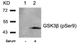
Western blot analysis of lysed extracts from 293 cells untreated (Lane 1) or treated with serum (lane 2) using GSK3β (Phospho-Ser9).IHC
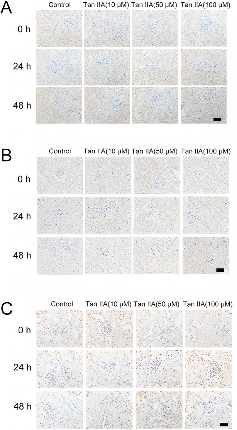
Tan IIA increased the expression of p-MEK (A), p-ERK1/2 (B) and p-GSK-3β (C), as assessed by immunohistochemical analysis.WB
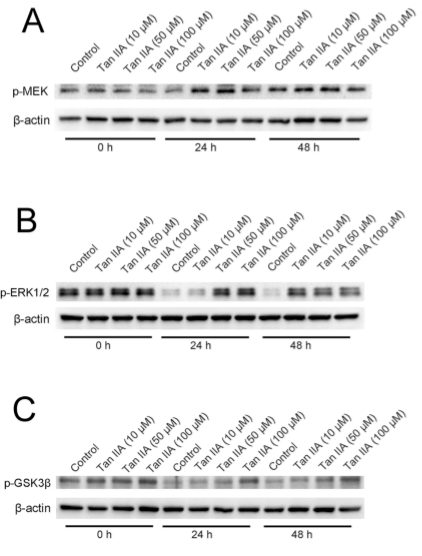
Tan IIA increases the expression of p-MEK (A), p-ERK1/2 (B) and p-GSK-3β (C), as detected by immunoblotting.WB
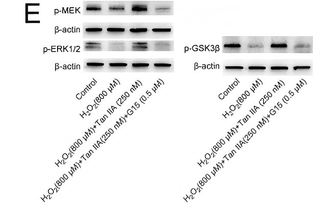
(E) Tan IIA increased the expression of p-MEK, p-ERK1/2 and p-GSK-3β, as detected by immunoblotting. G15 could diminish the expression of these proteins induced by Tan IIA. Previous| Product Name | GSK3beta (phospho Ser9) Antibody |
|---|---|
| Antibody Type | Primary Antibodies |
| Product description | Participates in the Wnt signaling pathway. Implicated in the hormonal control of several regulatory proteins including glycogen synthase, MYB and the transcription factor JUN. Phosphorylates JUN at sites proximal to its DNA-binding domain, thereby reducing its affinity for DNA. Phosphorylates MUC1 in breast cancer cells, and decreases the interaction of MUC1 with CTNNB1/beta-catenin. Phosphorylates CTNNB1/beta-catenin.1) Fan G, et al. (2003) J Biol Chem. 278 (52): 52432-52436. Barry FA, et al. (2003) FEBS Lett. 553 (1-2): 173-178. Welsh, et al. (1996) Trends Cell Biol. 6: 274-279. Srivastava A K, et al. (1998) Mol Cell Biochem. 182: 135-141. |
| Immunogen | GSK3beta (Phospho-Ser9) antibody was raised against a peptide sequence around phosphorylation site of serine 9 (T-T-S (p) -F-A) derived from Human GSK3β. |
| Modification | p-Ser9 |
| Clonality | Polyclonal |
|---|---|
| Host Species | Rabbit |
| Tested Applications | IFIHCWB |
| Western Blot: 1:500~1:1000, Immunohistochemistry: 1:50~1:100, Immunofluorescence: 1:100~1:200: |
|
| Species Reactivity | HumanMouseRat |
| Concentration | 1mg/ml |
| Purification | Affinity purified |
| Gene Symbol | GSK3B |
|---|---|
| Alternative Names | NFKB3 Factor A GSK-3 beta Protein kinase GSK-3-beta kinase GSK-3 beta |
| Molecular Weight(MW) | 46 kDa |
| Tissue Specificity | This antibody detects endogenous level of GSK3β only when phosphorylated at serine 9. |
| Entrez Gene | 2932 |
|---|---|
| Protein Accession | NP_001139628.1 |
Application
Top Image: Immunohistochemical analysis of paraffin-embedded human breast carcinoma tissue using GSK3β (Phospho-Ser9). Bottom Image: Immunofluorescence staining of methanol-fixed HeLa cells showing cytoplasmic staining using GSK3β (Phospho-Ser9).
Application
Western blot analysis of lysed extracts from 293 cells untreated (Lane 1) or treated with serum (lane 2) using GSK3β (Phospho-Ser9).
IHC
Tan IIA increased the expression of p-MEK (A), p-ERK1/2 (B) and p-GSK-3β (C), as assessed by immunohistochemical analysis.
WB
Tan IIA increases the expression of p-MEK (A), p-ERK1/2 (B) and p-GSK-3β (C), as detected by immunoblotting.
WB
(E) Tan IIA increased the expression of p-MEK, p-ERK1/2 and p-GSK-3β, as detected by immunoblotting. G15 could diminish the expression of these proteins induced by Tan IIA. Previous| Application Notes | Western Blot: 1:500~1:1000, Immunohistochemistry: 1:50~1:100, Immunofluorescence: 1:100~1:200: |
|---|
| Form | Liquid |
|---|---|
| Storage Instructions | Store antibody at -20˚C for up to one year. |
| Storage Buffer | Antibody supplied in phosphate buffered saline (without Mg2+ and Ca2+), pH 7.4, 150mM NaCl, 0.02% sodium azide and 50% glycerol. |


