Application
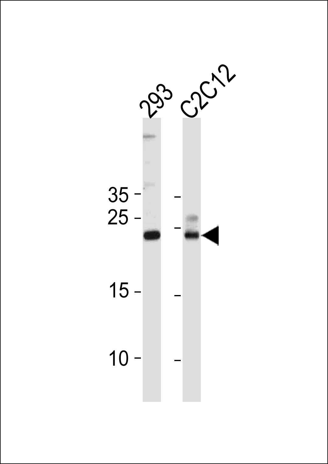
Western blot analysis in 293,mouse C2C12 cell line lysates (35ug/lane).Application
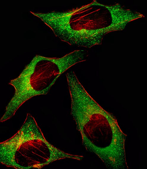
Fluorescent image of Hela cell stained with HIP2 Antibody .Hela cells were fixed with 4% PFA (20 min), permeabilized with Triton X-100 (0.1%, 10 min), then incubated with HIP2 primary antibody (1:25). For secondary antibody, Alexa Fluor 488 conjugated donkey anti-rabbit antibody (green) was used (1:400).Cytoplasmic actin was counterstained with Alexa Fluor 555 (red) conjugated Phalloidin (7units/mApplication
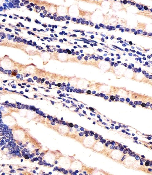
Immunohistochemical analysis of paraffin-embedded H. small intestine section using HIP2 Antibody . Antibody was diluted at 1:100 dilution. A undiluted biotinylated goat polyvalent antibody was used as the secondary, followed by DAB staining.| Product Name | HIP2 Antibody |
|---|---|
| Antibody Type | Primary Antibodies |
| Product description | HIP2 belongs to the ubiquitin-conjugating enzyme family. It binds selectively to a large region at the N terminus of huntingtin. This interaction is not influenced by the length of the huntingtin polyglutamine tract. This protein has been implicated in the degradation of huntingtin and suppression of apoptosis.1) Furukawa, Y., et al., Electrophoresis 21(2):338-346 (2000). |
| Immunogen | This HIP2 antibody is generated from rabbits immunized with a KLH conjugated synthetic peptide between 109-139 amino acids from the Central region of human HIP2. |
| Clonality | Polyclonal |
|---|---|
| Isotype | Ig |
| Host Species | Rabbit |
| Tested Applications | IFIHC-PWB |
| For IHC-P starting dilution is: 1:100 For IF starting dilution is: 1:10~50 For WB starting dilution is: 1:1000 | |
| Species Reactivity | HumanMouse |
| Concentration | 1mg/ml |
| Purification | Unpurified |
| Gene Symbol | UBE2K |
|---|---|
| Alternative Names | Ubiquitin-conjugating enzyme E2 K Huntingtin-interacting protein 2 HIP-2 Ubiquitin carrier protein Ubiquitin-conjugating enzyme E2-25 kDa Ubiquitin-conjugating enzyme E2(25K) Ubiquitin-conjugating enzyme E2-25K Ubiquitin-protein ligase UBE2K HIP2 |
| Molecular Weight(MW) | 22 kDa |
| Sequence Similarities | Predicted species reactivity based on immunogen sequence: Bovine |
Application
Western blot analysis in 293,mouse C2C12 cell line lysates (35ug/lane).
Application
Fluorescent image of Hela cell stained with HIP2 Antibody .Hela cells were fixed with 4% PFA (20 min), permeabilized with Triton X-100 (0.1%, 10 min), then incubated with HIP2 primary antibody (1:25). For secondary antibody, Alexa Fluor 488 conjugated donkey anti-rabbit antibody (green) was used (1:400).Cytoplasmic actin was counterstained with Alexa Fluor 555 (red) conjugated Phalloidin (7units/m
Application
Immunohistochemical analysis of paraffin-embedded H. small intestine section using HIP2 Antibody . Antibody was diluted at 1:100 dilution. A undiluted biotinylated goat polyvalent antibody was used as the secondary, followed by DAB staining.| Application Notes | For IHC-P starting dilution is: 1:100 For IF starting dilution is: 1:10~50 For WB starting dilution is: 1:1000 |
|---|
| Form | Liquid |
|---|---|
| Storage Instructions | Store at 4˚C for three months and -20˚C, stable for up to one year. As with all antibodies care should be taken to avoid repeated freeze thaw cycles. Antibodies should not be exposed to prolonged high temperatures. |
| Storage Buffer | Supplied in PBS with 0.09% (W/V) sodium azide. |


