Application
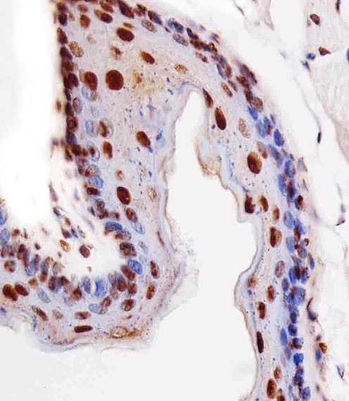
Immunohistochemical analysis of paraffin-embedded M. esophagus section using HIST1H4A Antibody . Antibody was diluted at 1:25 dilution. A peroxidase-conjugated goat anti-rabbit IgG at 1:400 dilution was used as the secondary antibody, followed by DAB staining.Application
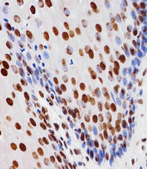
Immunohistochemical analysis of paraffin-embedded H. esophagus section using HIST1H4A Antibody . Antibody was diluted at 1:25 dilution. A peroxidase-conjugated goat anti-rabbit IgG at 1:400 dilution was used as the secondary antibody, followed by DAB staining.Application
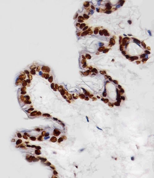
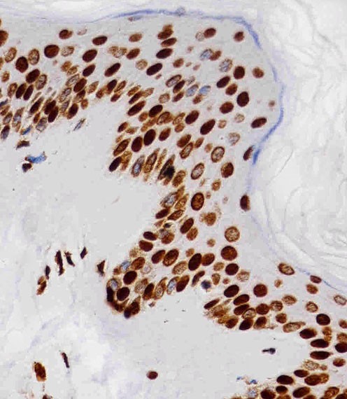
Immunohistochemical analysis of paraffin-embedded M. skin section using HIST1H4A Antibody . Antibody was diluted at 1:25 dilution. A peroxidase-conjugated goat anti-rabbit IgG at 1:400 dilution was used as the secondary antibody, followed by DAB staining.Application
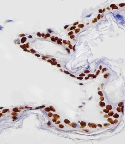
Immunohistochemical analysis of paraffin-embedded R. skin section using HIST1H4A Antibody . Antibody was diluted at 1:25 dilution. A peroxidase-conjugated goat anti-rabbit IgG at 1:400 dilution was used as the secondary antibody, followed by DAB staining.Application
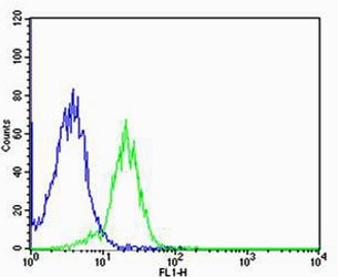
Flow cytometric analysis of MCF-7 cells using HIST1H4A Antibody (green) compared to an isotype control of rabbit IgG(blue). Antibody was diluted at 1:25 dilution. An Alexa Fluor 488 goat anti-rabbit lgG at 1:400 dilution was used as the secondary antibody.Application
Immunohistochemical analysis of paraffin-embedded H. skin section using HIST1H4A Antibody . Antibody was diluted at 1:25 dilution. A peroxidase-conjugated goat anti-rabbit IgG at 1:400 dilution was used as the secondary antibody, followed by DAB staining.Application
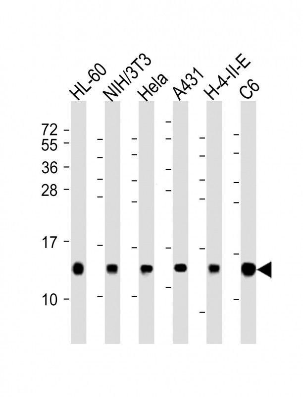
Western Blot at 1:2000 dilution Lane 1: HL-60 whole cell lysates Lane 2: NIH/3T3 whole cell lysates Lane 3: Hela whole cell lysates Lane 4: A431 whole cell lysates Lane 5: H-4-Ⅱ-E whole cell lysates Lane 6: C6 whole cell lysates Lysates/proteins at 20 ug per lane.| Product Name | HIST1H4A Antibody |
|---|---|
| Antibody Type | Primary Antibodies |
| Antigen Alias | Histone H4, HIST1H4A, H4/A, H4FA |
| Product description | Core component of nucleosome. Nucleosomes wrap and compact DNA into chromatin, limiting DNA accessibility to the cellular machineries which require DNA as a template. Histones thereby play a central role in transcription regulation, DNA repair, DNA replication and chromosomal stability. DNA accessibility is regulated via a complex set of post-translational modifications of histones, also called histone code, and nucleosome remodeling.1) Sierra F.,et al.Nucleic Acids Res. 11:7069-7086(1983). |
| Immunogen | This HIST1H4A antibody is generated from a rabbit immunized with a KLH conjugated synthetic peptide between 71-103 amino acids from the C-terminal region of human HIST1H4A. |
| Clonality | Polyclonal |
|---|---|
| Isotype | Ig |
| Host Species | Rabbit |
| Tested Applications | FACSIHC-PWB |
| For WB starting dilution is: 1:2000 For IHC-P starting dilution is: 1:25 For FACS starting dilution is: 1:25: |
|
| Species Reactivity | HumanMouseRat |
| Concentration | 1mg/ml |
| Purification | Affinity purified |
| Gene Symbol | HIST1H4A |
|---|---|
| Alternative Names | Histone H4 HIST1H4A H4/A H4FA |
| Molecular Weight(MW) | 11 kDa |
| Sequence Similarities | Predicted species reactivity based on immunogen sequence: Bovine |
| Entrez Gene | 121504 554313 8294 8359 8360 8361 8362 8363 8364 8365 8366 8367 8368 8370 |
|---|---|
| Protein Accession | P62805 |
Application
Immunohistochemical analysis of paraffin-embedded M. esophagus section using HIST1H4A Antibody . Antibody was diluted at 1:25 dilution. A peroxidase-conjugated goat anti-rabbit IgG at 1:400 dilution was used as the secondary antibody, followed by DAB staining.
Application
Immunohistochemical analysis of paraffin-embedded H. esophagus section using HIST1H4A Antibody . Antibody was diluted at 1:25 dilution. A peroxidase-conjugated goat anti-rabbit IgG at 1:400 dilution was used as the secondary antibody, followed by DAB staining.
Application
Immunohistochemical analysis of paraffin-embedded M. skin section using HIST1H4A Antibody . Antibody was diluted at 1:25 dilution. A peroxidase-conjugated goat anti-rabbit IgG at 1:400 dilution was used as the secondary antibody, followed by DAB staining.
Application
Immunohistochemical analysis of paraffin-embedded R. skin section using HIST1H4A Antibody . Antibody was diluted at 1:25 dilution. A peroxidase-conjugated goat anti-rabbit IgG at 1:400 dilution was used as the secondary antibody, followed by DAB staining.
Application
Flow cytometric analysis of MCF-7 cells using HIST1H4A Antibody (green) compared to an isotype control of rabbit IgG(blue). Antibody was diluted at 1:25 dilution. An Alexa Fluor 488 goat anti-rabbit lgG at 1:400 dilution was used as the secondary antibody.
Application
Immunohistochemical analysis of paraffin-embedded H. skin section using HIST1H4A Antibody . Antibody was diluted at 1:25 dilution. A peroxidase-conjugated goat anti-rabbit IgG at 1:400 dilution was used as the secondary antibody, followed by DAB staining.
Application
Western Blot at 1:2000 dilution Lane 1: HL-60 whole cell lysates Lane 2: NIH/3T3 whole cell lysates Lane 3: Hela whole cell lysates Lane 4: A431 whole cell lysates Lane 5: H-4-Ⅱ-E whole cell lysates Lane 6: C6 whole cell lysates Lysates/proteins at 20 ug per lane.| Application Notes | For WB starting dilution is: 1:2000 For IHC-P starting dilution is: 1:25 For FACS starting dilution is: 1:25: |
|---|
| Form | Liquid |
|---|---|
| Storage Instructions | Store at 4˚C for three months and -20˚C, stable for up to one year. As with all antibodies care should be taken to avoid repeated freeze thaw cycles. Antibodies should not be exposed to prolonged high temperatures. |
| Storage Buffer | Supplied in PBS with 0.09% (W/V) sodium azide. |


