Application
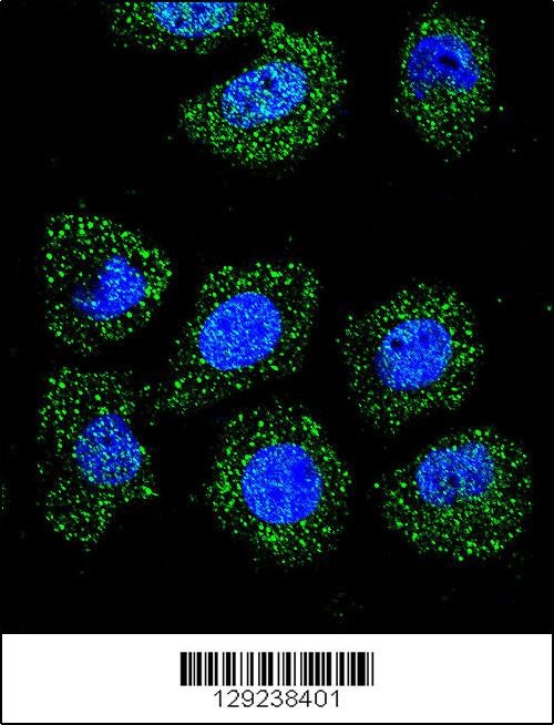
Confocal immunofluorescent analysis of ID1 Antibody with U-251MG cell followed by Alexa Fluor 488-conjugated goat anti-rabbit lgG (green).DAPI was used to stain the cell nuclear (blue).Application
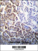
ID1 Antibody immunohistochemistry analysis in formalin fixed and paraffin embedded human pancreas tissue followed by peroxidase conjugation of the secondary antibody and DAB staining.Application
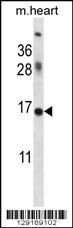
Western blot analysis in mouse heart tissue lysates (35ug/lane).This demonstrates the ID1(PEI 1:1detected the ID1 protein (arrow).Application
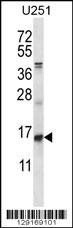
Western blot analysis in U251 cell line lysates (35ug/lane).Application
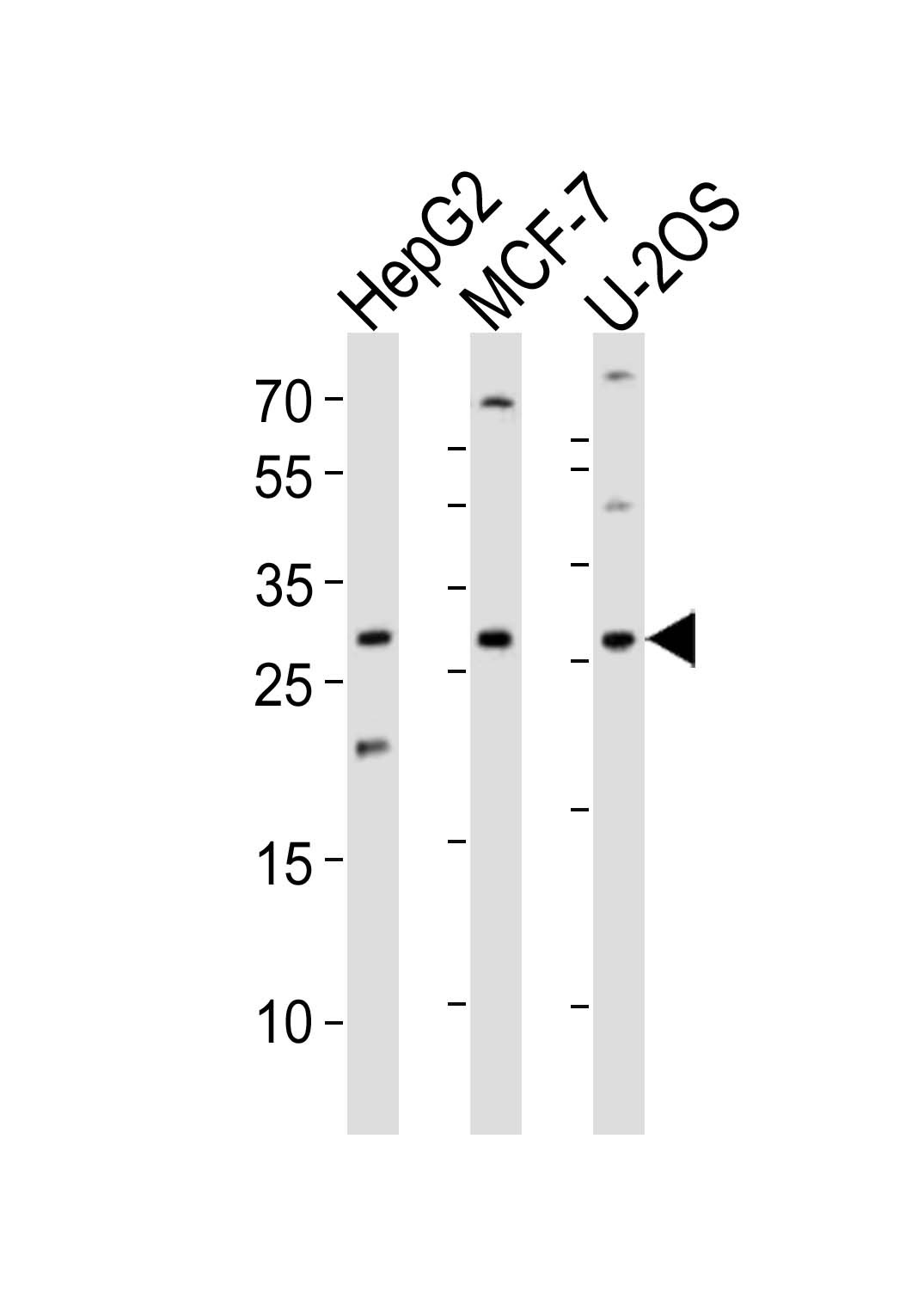
Western blot analysis of lysates from HepG2, MCF-7, U-2OS cell line (from left to right), using ID1 Antibody at 1:1000 at each lane.Application
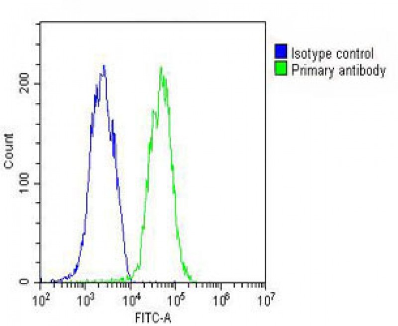
Overlay histogram showing HepG2 cells stained with Antibody (green line). The cells were fixed with 2% paraformaldehyde (10 min) and then permeabilized with 90% methanol for 10 min. The cells were then icubated in 2% bovine serum albumin to block non-specific protein-protein interactions followed by the antibody (1:25 dilution) for 60 min at 37ºC. The secondary antibody used was Goat-Anti-Rabbit I| Product Name | ID1 Antibody |
|---|---|
| Antibody Type | Primary Antibodies |
| Antigen Alias | DNA-binding protein inhibitor ID-1, Class B basic helix-loop-helix protein 24, bHLHb24, Inhibitor of DNA binding 1, Inhibitor of differentiation 1, ID1, BHLHB24, ID |
| Product description | The protein encoded by this gene is a helix-loop-helix (HLH) protein that can form heterodimers with members of the basic HLH family of transcription factors. The encoded protein has no DNA binding activity and therefore can inhibit the DNA binding and transcriptional activation ability of basic HLH proteins with which it interacts. This protein may play a role in cell growth, senescence, and differentiation. Two transcript variants encoding different isoforms have been found for this gene. [provided by RefSeq].1) Qian, T., et al. Oncogene 29(43):5818-5827(2010) |
| Immunogen | This ID1 antibody is generated from rabbits immunized with a KLH conjugated synthetic peptide between 66-93 amino acids from the Central region of human ID1. |
| Clonality | Polyclonal |
|---|---|
| Isotype | Ig |
| Host Species | Rabbit |
| Tested Applications | FACSIFIHC-PWB |
| For FACS starting dilution is: 1:25 For WB starting dilution is: 1:1000 For IHC-P starting dilution is: 1:10~50 For IF starting dilution is: 1:10~50: |
|
| Species Reactivity | HumanRat |
| Concentration | 1mg/ml |
| Purification | Affinity purified |
| Gene Symbol | ID1 |
|---|---|
| Alternative Names | DNA-binding protein inhibitor ID-1 Class B basic helix-loop-helix protein 24 bHLHb24 Inhibitor of DNA binding 1 Inhibitor of differentiation 1 ID1 BHLHB24 ID |
| Molecular Weight(MW) | 16 kDa |
Application
Confocal immunofluorescent analysis of ID1 Antibody with U-251MG cell followed by Alexa Fluor 488-conjugated goat anti-rabbit lgG (green).DAPI was used to stain the cell nuclear (blue).
Application
ID1 Antibody immunohistochemistry analysis in formalin fixed and paraffin embedded human pancreas tissue followed by peroxidase conjugation of the secondary antibody and DAB staining.
Application
Western blot analysis in mouse heart tissue lysates (35ug/lane).This demonstrates the ID1(PEI 1:1detected the ID1 protein (arrow).
Application
Western blot analysis in U251 cell line lysates (35ug/lane).
Application
Western blot analysis of lysates from HepG2, MCF-7, U-2OS cell line (from left to right), using ID1 Antibody at 1:1000 at each lane.
Application
Overlay histogram showing HepG2 cells stained with Antibody (green line). The cells were fixed with 2% paraformaldehyde (10 min) and then permeabilized with 90% methanol for 10 min. The cells were then icubated in 2% bovine serum albumin to block non-specific protein-protein interactions followed by the antibody (1:25 dilution) for 60 min at 37ºC. The secondary antibody used was Goat-Anti-Rabbit I| Application Notes | For FACS starting dilution is: 1:25 For WB starting dilution is: 1:1000 For IHC-P starting dilution is: 1:10~50 For IF starting dilution is: 1:10~50: |
|---|
| Form | Liquid |
|---|---|
| Storage Instructions | Store at 4˚C for three months and -20˚C, stable for up to one year. As with all antibodies care should be taken to avoid repeated freeze thaw cycles. Antibodies should not be exposed to prolonged high temperatures. |
| Storage Buffer | Supplied in PBS with 0.09% (W/V) sodium azide. |


