Application
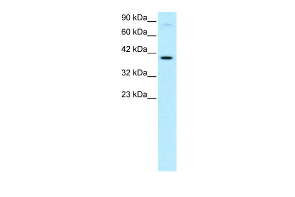
Antibody used in WB on Human HepG2 at 0.2-1 ug/ml.Application
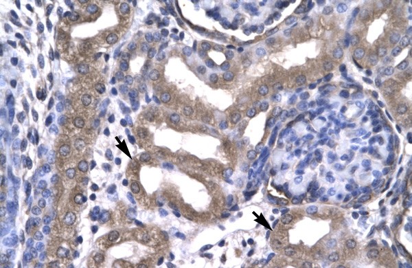
Antibody used in IHC on Human kidney lysate.Application
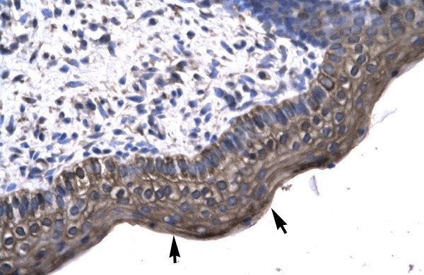
Antibody used in IHC on Human Spermatophore.| Product Name | KIN Antibody |
|---|---|
| Antibody Type | Primary Antibodies |
| Product description | Kin is a nuclear protein that forms intranuclear foci during proliferation and is redistributed in the nucleoplasm during the cell cycle. Short-wave ultraviolet light provokes the relocalization of the protein, suggesting its participation in the cellular response to DNA damage. Originally selected based on protein-binding with RecA antibodies, the mouse protein presents a limited similarity with a functional domain of the bacterial RecA protein, a characteristic shared by the human ortholog.1) Despras, E., et al., (2003) Radiat.Res.159(6), 748-758. |
| Immunogen | Antibody produced in rabbits immunized with a synthetic peptide corresponding a region of human KIN. |
| Clonality | Polyclonal |
|---|---|
| Host Species | Rabbit |
| Tested Applications | ELISAIHCWB |
| KIN antibody can be used for detection of KIN by ELISA at 1:2500. KIN antibody can be used for detection of KIN by western blot at 0.2-5.0 μg/mL, and HRP conjugated secondary antibody should be diluted 1:50,000 - 100,000. | |
| Species Reactivity | Human |
| Concentration | 1mg/ml |
| Purification | Affinity purified |
| Gene Symbol | KIN |
|---|---|
| Alternative Names | KIN BTCD KIN17 |
| Molecular Weight(MW) | 45 kDa |
Application
Antibody used in WB on Human HepG2 at 0.2-1 ug/ml.
Application
Antibody used in IHC on Human kidney lysate.
Application
Antibody used in IHC on Human Spermatophore.| Positive Control | 1) Cat. No. 1211 - HepG2 Cell Lysate |
|---|---|
| Application Notes | KIN antibody can be used for detection of KIN by ELISA at 1:2500. KIN antibody can be used for detection of KIN by western blot at 0.2-5.0 μg/mL, and HRP conjugated secondary antibody should be diluted 1:50,000 - 100,000. |
| Form | Liquid |
|---|---|
| Storage Instructions | For short periods of storage (days) store at 4˚C. For longer periods of storage, store KIN antibody at -20˚C. As with any antibody avoid repeat freeze-thaw cycles. |
| Storage Buffer | Antibody is lyophilized in PBS buffer with 2% sucrose. Add 50 μL of distilled water. Final antibody concentration is 1 mg/mL. |


