Application
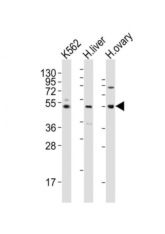
Western Blot at 1:2000 dilution Lane 1: K562 whole cell lysates Lane 2: human liver lysates Lane 3: human ovary lysates Lysates/proteins at 20 ug per lane.Application
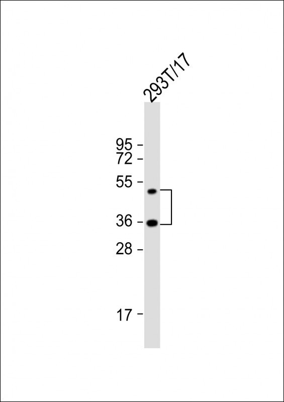
Western Blot at 1:2000 dilution + 293T/17 whole cell lysate Lysates/proteins at 20 ug per lane.Application
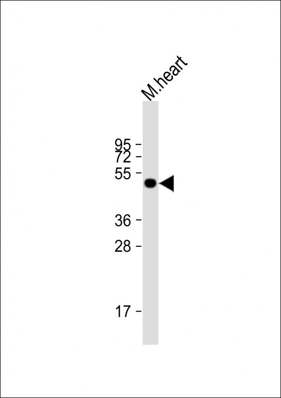
Western Blot at 1:2000 dilution + mouse heart lysate Lysates/proteins at 20 ug per lane.Application
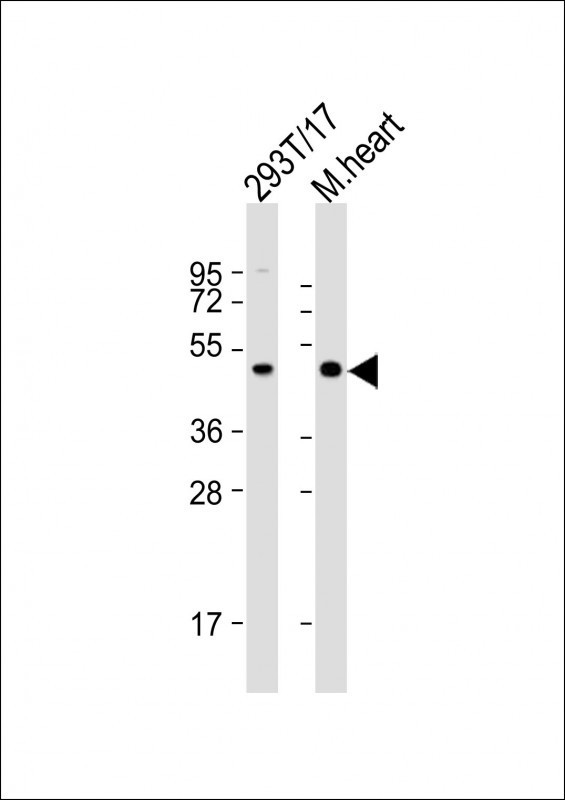
Western Blot at 1:2000 dilution Lane 1: 293T/17 whole cell lysate Lane 2: mouse heart lysate Lysates/proteins at 20 ug per lane.Application
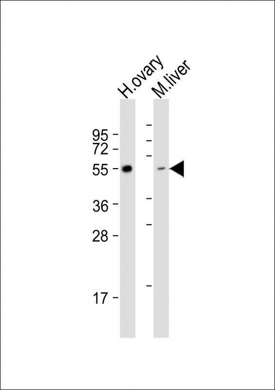
Western Blot at 1:1000 dilution Lane 1: human ovary lysate Lane 2: mouse liver lysate Lysates/proteins at 20 ug per lane.Application
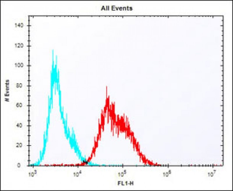
Overlay histogram showing Jurkat cells stained with Antibody (red line). The cells were fixed with 2% paraformaldehyde (10 min) and then permeabilized with 90% methanol for 10 min. The cells were then icubated in 2% bovine serum albumin to block non-specific protein-protein interactions followed by the antibody (1:25 dilution) for 60 min at 37ºC. The secondary antibody used was Alexa Fluor 488 goaApplication
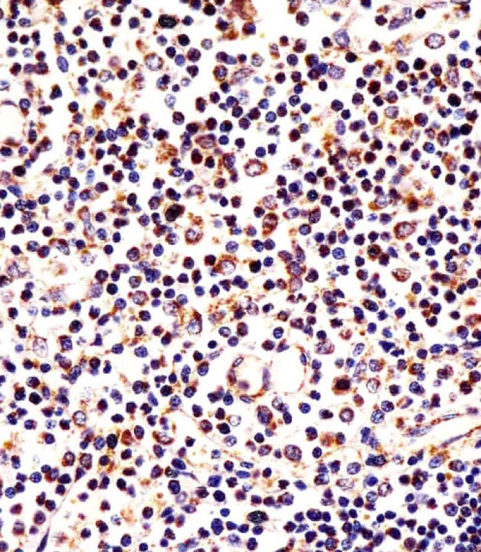
Antibody staining LAG3 in human thymus tissue sections by Immunohistochemistry (IHC-P - paraformaldehyde-fixed, paraffin-embedded sections).| Product Name | LAG3 Antibody |
|---|---|
| Antibody Type | Primary Antibodies |
| Antigen Alias | Lymphocyte activation gene 3 protein, LAG-3, Protein FDC, CD223, LAG3, FDC |
| Product description | Lymphocyte-activation protein 3 belongs to Ig superfamily and contains 4 extracellular Ig-like domains.1) Smyth,D.J., et.al., BMC Med. Genet. 7, 20 (2006) |
| Immunogen | This LAG3 antibody is generated from rabbits immunized with a KLH conjugated synthetic peptide between 103-132 amino acids from the Central region of human LAG3. |
| Clonality | Polyclonal |
|---|---|
| Isotype | Ig |
| Host Species | Rabbit |
| Tested Applications | IHC-P |
| For IHC-P starting dilution is: 1:25 For FACS starting dilution is: 1:25 For WB starting dilution is: 1:1000: |
|
| Species Reactivity | HumanMouse |
| Concentration | 1mg/ml |
| Purification | Affinity purified |
| Gene Symbol | LAG3 |
|---|---|
| Alternative Names | Lymphocyte activation gene 3 protein LAG-3 Protein FDC CD223 LAG3 FDC |
| Molecular Weight(MW) | 57 kDa |
Application
Western Blot at 1:2000 dilution Lane 1: K562 whole cell lysates Lane 2: human liver lysates Lane 3: human ovary lysates Lysates/proteins at 20 ug per lane.
Application
Western Blot at 1:2000 dilution + 293T/17 whole cell lysate Lysates/proteins at 20 ug per lane.
Application
Western Blot at 1:2000 dilution + mouse heart lysate Lysates/proteins at 20 ug per lane.
Application
Western Blot at 1:2000 dilution Lane 1: 293T/17 whole cell lysate Lane 2: mouse heart lysate Lysates/proteins at 20 ug per lane.
Application
Western Blot at 1:1000 dilution Lane 1: human ovary lysate Lane 2: mouse liver lysate Lysates/proteins at 20 ug per lane.
Application
Overlay histogram showing Jurkat cells stained with Antibody (red line). The cells were fixed with 2% paraformaldehyde (10 min) and then permeabilized with 90% methanol for 10 min. The cells were then icubated in 2% bovine serum albumin to block non-specific protein-protein interactions followed by the antibody (1:25 dilution) for 60 min at 37ºC. The secondary antibody used was Alexa Fluor 488 goa
Application
Antibody staining LAG3 in human thymus tissue sections by Immunohistochemistry (IHC-P - paraformaldehyde-fixed, paraffin-embedded sections).| Application Notes | For IHC-P starting dilution is: 1:25 For FACS starting dilution is: 1:25 For WB starting dilution is: 1:1000: |
|---|
| Form | Liquid |
|---|---|
| Storage Instructions | Store at 4˚C for three months and -20˚C, stable for up to one year. As with all antibodies care should be taken to avoid repeated freeze thaw cycles. Antibodies should not be exposed to prolonged high temperatures. |
| Storage Buffer | Supplied in PBS with 0.09% (W/V) sodium azide. |


 Data sheet for OM283198