Application
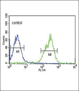
Flow cytometric analysis of HepG2 cells (right histogram) compared to a negative control cell (left histogram).FITC-conjugated goat-anti-rabbit secondary antibodies were used for the analysis.Application
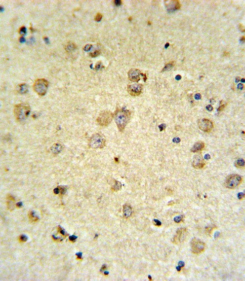
MAP4K3 Antibody IHC analysis in formalin fixed and paraffin embedded brain tissue followed by peroxidase conjugation of the secondary antibody and DAB staining.Application
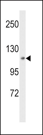
Western blot analysis of MAP4K3 Antibody in HepG2 cell line lysates (35ug/lane)| Product Name | MAP4K3 Antibody |
|---|---|
| Antibody Type | Primary Antibodies |
| Product description | MAP4K3 encodes a member of the Ste20 family of serine/threonine protein kinases. The protein belongs to the subfamily that consists of members, such as germinal center kinase (GCK), that are characterized by an N-terminal catalytic domain and C-terminal regulatory domain. The kinase activity of the encoded protein can be stimulated by UV radiation and tumor necrosis factor-alpha. The protein specifically activates the c-Jun N-terminal kinase (JNK) signaling pathway. Evidence suggests that it functions upstream of mitogen-activated protein kinase kinase kinase 1 (MEKK1). This gene previously was referred to as RAB8-interacting protein-like 1 (RAB8IPL1), but it has been renamed mitogen-activated protein kinase kinase kinase kinase 3 (MAP4K3).1) Yan, L., et al. Mol. Cell 37(5):633-642(2010) |
| Immunogen | This MAP4K3 antibody is generated from rabbits immunized with a recombinant protein of human MAP4K3. |
| Clonality | Polyclonal |
|---|---|
| Isotype | Ig |
| Host Species | Rabbit |
| Tested Applications | FACSIHC-PWB |
| For WB starting dilution is: 1:1000 For IHC-P starting dilution is: 1:10~50 For FACS starting dilution is: 1:10~50 | |
| Species Reactivity | Human |
| Concentration | 1mg/ml |
| Purification | Affinity purified |
| Gene Symbol | MAP4K3 |
|---|---|
| Alternative Names | Mitogen-activated protein kinase kinase kinase kinase 3 Germinal center kinase-related protein kinase GLK MAPK/ERK kinase kinase kinase 3 MEK kinase kinase 3 MEKKK 3 MAP4K3 RAB8IPL1 |
| Molecular Weight(MW) | 101 kDa |
Application
Flow cytometric analysis of HepG2 cells (right histogram) compared to a negative control cell (left histogram).FITC-conjugated goat-anti-rabbit secondary antibodies were used for the analysis.
Application
MAP4K3 Antibody IHC analysis in formalin fixed and paraffin embedded brain tissue followed by peroxidase conjugation of the secondary antibody and DAB staining.
Application
Western blot analysis of MAP4K3 Antibody in HepG2 cell line lysates (35ug/lane)| Application Notes | For WB starting dilution is: 1:1000 For IHC-P starting dilution is: 1:10~50 For FACS starting dilution is: 1:10~50 |
|---|
| Form | Liquid |
|---|---|
| Storage Instructions | Store at 4˚C for three months and -20˚C, stable for up to one year. As with all antibodies care should be taken to avoid repeated freeze thaw cycles. Antibodies should not be exposed to prolonged high temperatures. |
| Storage Buffer | Supplied in PBS with 0.09% (W/V) sodium azide. |


