Application
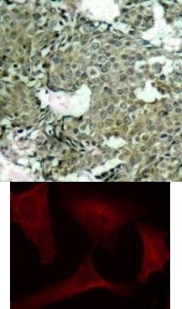
Top Image: Immunohistochemical analysis of paraffin-embedded human breast carcinoma tissue using MEK1 (Phospho-Ser221). Bottom Image: Immunofluorescence staining of methanol-fixed HeLa cells using MEK1 (Phospho-Ser221).Application
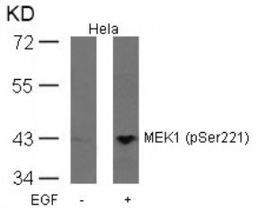
Western blot analysis of lysed extracts from HeLa cells untreated or treated with EGF using MEK1 (Phospho-Ser221).IHC
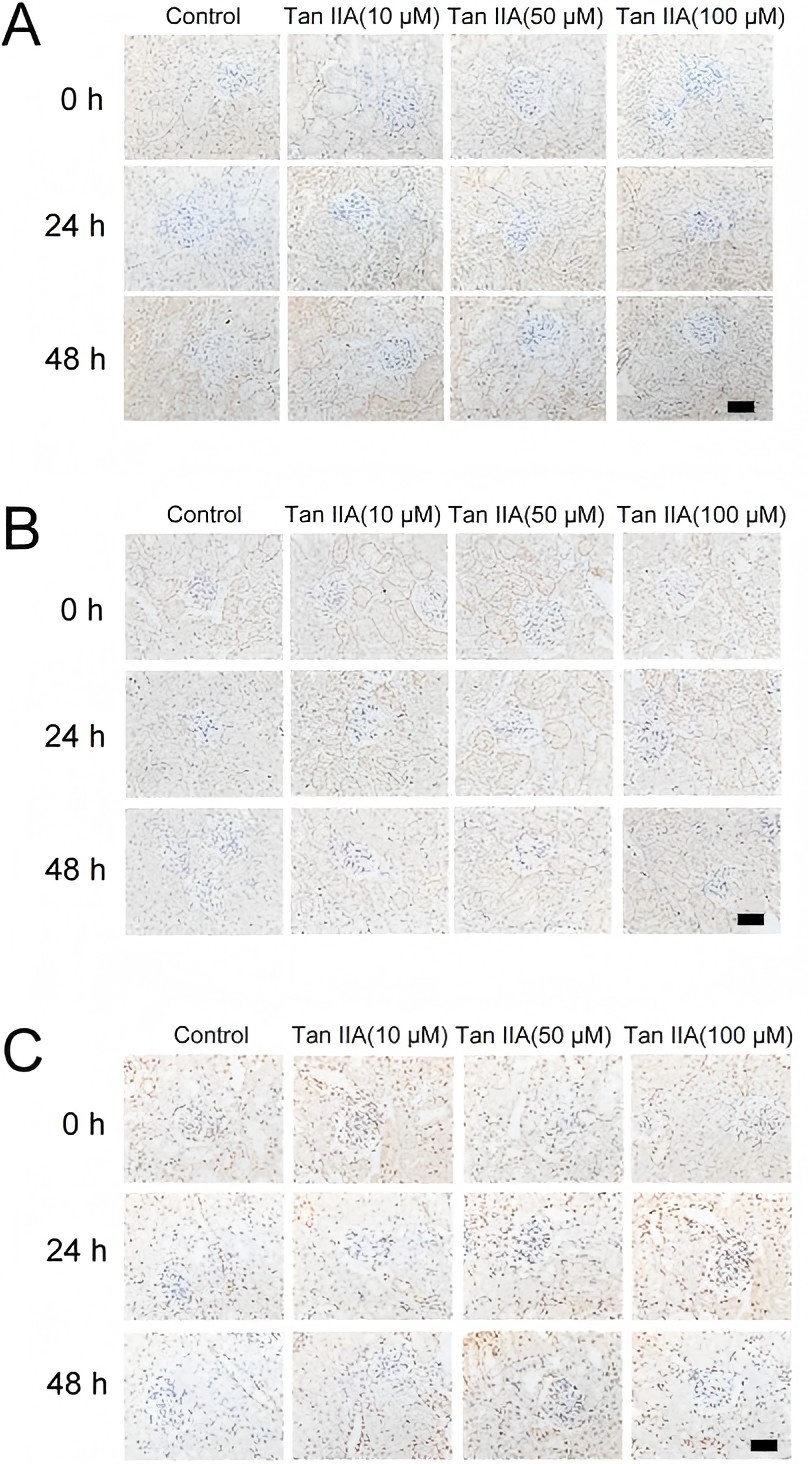
Tan IIA increased the expression of p-MEK (A), p-ERK1/2 (B) and p-GSK-3β (C), as assessed by immunohistochemical analysis.WB
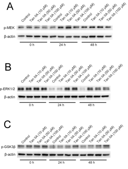
Tan IIA increases the expression of p-MEK (A), p-ERK1/2 (B) and p-GSK-3β (C), as detected by immunoblotting.WB
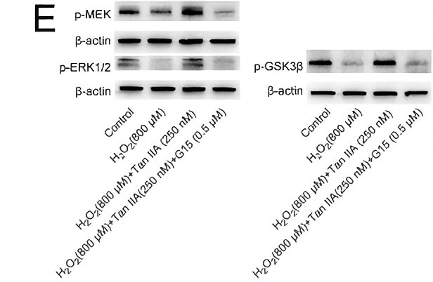
(E) Tan IIA increased the expression of p-MEK, p-ERK1/2 and p-GSK-3β, as detected by immunoblotting. G15 could diminish the expression of these proteins induced by Tan IIA. Previous| Product Name | MEK1 (phospho Ser221) Antibody |
|---|---|
| Antibody Type | Primary Antibodies |
| Product description | Catalyzes the concomitant phosphorylation of a threonine and a tyrosine residue in a Thr-Glu-Tyr sequence located in MAP kinases. Activates ERK1 and ERK2 MAP kinases.1) Zebisch A, et al. (2006) Cancer Res; 66 (7): 3401-8. Luciano BS, et al. (2006) J Biol Chem; 279 (50): 52117-23. Wang X, et al. (2003) Oncogene; 22 (1): 109-16. Gopalbhai K, et al. (2003) J Biol Chem; 278 (10): 8118-25. Ling MT, et al. (2002) Oncogene; 21 (55): 8498-505. |
| Immunogen | MEK1 (Phospho-Ser221) antibody was raised against a peptide sequence around phosphorylation site of serine 221 (A-N-S (p) -F-V) derived from Human MEK1. |
| Modification | p-Ser221 |
| Clonality | Polyclonal |
|---|---|
| Host Species | Rabbit |
| Tested Applications | IFIHCWB |
| Western Blot: 1:500~1:1000, Immunohistochemistry: 1:50~1:100, Immunofluorescence: 1:100~1:200 | |
| Species Reactivity | HumanMouseRat |
| Concentration | 1mg/ml |
| Purification | Affinity purified |
| Gene Symbol | MAP2K1 |
|---|---|
| Alternative Names | CFC3 MEK1 MKK1 MAPKK1 PRKMK1 EMS1 ERK activator kinase 1 MAP kinase kinase 1 MAP2K1 MAPK/ERK kinase 1 MAPKK 1 |
| Molecular Weight(MW) | 45 kDa |
| Tissue Specificity | This antibody detects endogenous level of MEK1 only when phosphorylated at serine 221. |
| Entrez Gene | 5604 |
|---|---|
| Protein Accession | NP_002746.1 |
Application
Top Image: Immunohistochemical analysis of paraffin-embedded human breast carcinoma tissue using MEK1 (Phospho-Ser221). Bottom Image: Immunofluorescence staining of methanol-fixed HeLa cells using MEK1 (Phospho-Ser221).
Application
Western blot analysis of lysed extracts from HeLa cells untreated or treated with EGF using MEK1 (Phospho-Ser221).
IHC
Tan IIA increased the expression of p-MEK (A), p-ERK1/2 (B) and p-GSK-3β (C), as assessed by immunohistochemical analysis.
WB
Tan IIA increases the expression of p-MEK (A), p-ERK1/2 (B) and p-GSK-3β (C), as detected by immunoblotting.
WB
(E) Tan IIA increased the expression of p-MEK, p-ERK1/2 and p-GSK-3β, as detected by immunoblotting. G15 could diminish the expression of these proteins induced by Tan IIA. Previous| Application Notes | Western Blot: 1:500~1:1000, Immunohistochemistry: 1:50~1:100, Immunofluorescence: 1:100~1:200 |
|---|
| Form | Liquid |
|---|---|
| Storage Instructions | Store antibody at -20˚C for up to one year. |
| Storage Buffer | Antibody supplied in phosphate buffered saline (without Mg2+ and Ca2+), pH 7.4, 150mM NaCl, 0.02% sodium azide and 50% glycerol. |


