Application
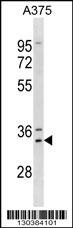
Western blot analysis in A375 cell line lysates (35ug/lane).Application
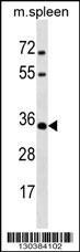
Western blot analysis in mouse spleen tissue lysates (35ug/lane).Application
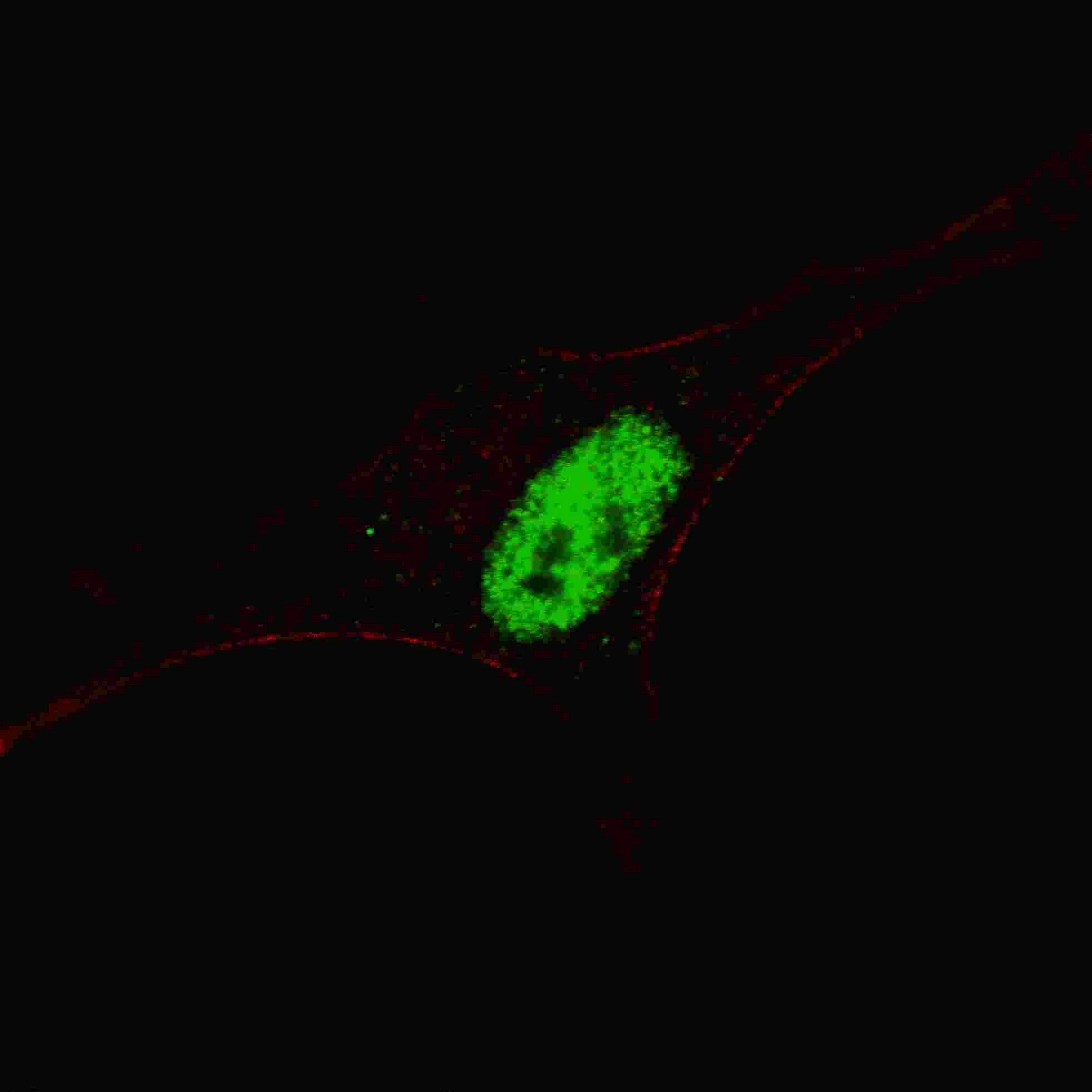
Fluorescent confocal image of SY5Y cells stained with MSX1 antibody. SY5Y cells were fixed with 4% PFA (20 min), permeabilized with Triton X-100 (0.2%, 30 min). Cells were then incubated with MSX1 primary antibody (1:200, 2 h at room temperature). For secondary antibody, Alexa Fluor 488 conjugated donkey anti-rabbit antibody (green) was used (1:1000, 1h). Nuclei were counterstained with Hoechst 33Application
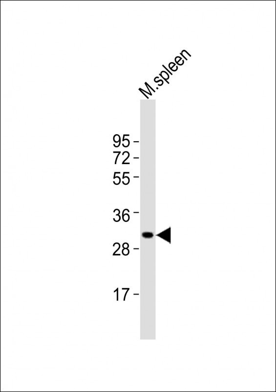
Western Blot at 1:2000 dilution + mouse spleen lysates Lysates/proteins at 20 ug per lane.Application
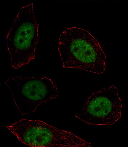
Fluorescent image of U251 cell stained with MSX1 Antibody .U251 cells were fixed with 4% PFA (20 min), permeabilized with Triton X-100 (0.1%, 10 min), then incubated with MSX1 primary antibody (1:25). For secondary antibody, Alexa Fluor 488 conjugated donkey anti-rabbit antibody (green) was used (1:400).Cytoplasmic actin was counterstained with Alexa Fluor 555 (red) conjugated Phalloidin (7units/m| Product Name | MSX1 Antibody |
|---|---|
| Antibody Type | Primary Antibodies |
| Antigen Alias | Homeobox protein MSX-1, Homeobox protein Hox-7, Msh homeobox 1-like protein, MSX1, HOX7 |
| Product description | This gene encodes a member of the muscle segment homeobox gene family. The encoded protein functions as a transcriptional repressor during embryogenesis through interactions with components of the core transcription complex and other homeoproteins. It may also have roles in limb-pattern formation, craniofacial development, particularly odontogenesis, and tumor growth inhibition. Mutations in this gene, which was once known as homeobox 7, have been associated with nonsyndromic cleft lip with or without cleft palate 5, Witkop syndrome, Wolf-Hirschom syndrome, and autosomoal dominant hypodontia.1) References for protein: |
| Immunogen | This MSX1 antibody is generated from rabbits immunized with a KLH conjugated synthetic peptide between 111-138 amino acids from the Central region of human MSX1. |
| Clonality | Polyclonal |
|---|---|
| Isotype | Ig |
| Host Species | Rabbit |
| Tested Applications | IFWB |
| For WB starting dilution is: 1:2000 For IF starting dilution is: 1:10~50: |
|
| Species Reactivity | HumanMouse |
| Concentration | 1mg/ml |
| Purification | Affinity purified |
| Gene Symbol | MSX1 |
|---|---|
| Alternative Names | Homeobox protein MSX-1 Homeobox protein Hox-7 Msh homeobox 1-like protein MSX1 HOX7 |
| Molecular Weight(MW) | 31 kDa |
| Sequence Similarities | Predicted species reactivity based on immunogen sequence: Bovine |
Application
Western blot analysis in A375 cell line lysates (35ug/lane).
Application
Western blot analysis in mouse spleen tissue lysates (35ug/lane).
Application
Fluorescent confocal image of SY5Y cells stained with MSX1 antibody. SY5Y cells were fixed with 4% PFA (20 min), permeabilized with Triton X-100 (0.2%, 30 min). Cells were then incubated with MSX1 primary antibody (1:200, 2 h at room temperature). For secondary antibody, Alexa Fluor 488 conjugated donkey anti-rabbit antibody (green) was used (1:1000, 1h). Nuclei were counterstained with Hoechst 33
Application
Western Blot at 1:2000 dilution + mouse spleen lysates Lysates/proteins at 20 ug per lane.
Application
Fluorescent image of U251 cell stained with MSX1 Antibody .U251 cells were fixed with 4% PFA (20 min), permeabilized with Triton X-100 (0.1%, 10 min), then incubated with MSX1 primary antibody (1:25). For secondary antibody, Alexa Fluor 488 conjugated donkey anti-rabbit antibody (green) was used (1:400).Cytoplasmic actin was counterstained with Alexa Fluor 555 (red) conjugated Phalloidin (7units/m| Application Notes | For WB starting dilution is: 1:2000 For IF starting dilution is: 1:10~50: |
|---|
| Form | Liquid |
|---|---|
| Storage Instructions | Store at 4˚C for three months and -20˚C, stable for up to one year. As with all antibodies care should be taken to avoid repeated freeze thaw cycles. Antibodies should not be exposed to prolonged high temperatures. |
| Storage Buffer | Supplied in PBS with 0.09% (W/V) sodium azide. |


