Application
NMDA NR2C Subunit Antibody| Product Name | NMDA NR2C Antibody |
|---|---|
| Antibody Type | Primary Antibodies |
| Antigen Alias | NR2C, GluN2C, N-methyl D-aspartate receptor subtype 2C |
| Product description | The ion channels activated by glutamate are typically divided into two classes. Those that are sensitive to N-methyl-D-aspartate (NMDA) are designated NMDA receptors (NMDAR). The NMDAR plays an essential role in memory, neuronal development and it has also been implicated in several disorders of the central nervous system including Alzheimer’s, epilepsy and ischemic neuronal cell death. The rat NMDAR1 (NR1) was the first subunit of the NMDAR to be cloned. The NR1 protein can form NMDA activated channels when expressed in Xenopus oocytes but the currents in such channels are much smaller than those seen in situ. Channels with more physiological characteristics are produced when the NR1 subunit is combined with one or more of the NMDAR2 (NR2 A-D) subunits.1) Thompson, C.L, Drewery, D.L., Atkins, H.D., Stephenson, F.A. and Chazot, P.L., "Immunohistochemical localization of N-methyl-D-aspartate receptor NR1, NR2A, NR2B and NR2C/D subunits in the adult mammalian cerebellum," Neurosci. Lett. 283 (2000) 85 - 88. |
| Immunogen | NMDA NR2C polyclonal antibody was raised against an N-terminal fusion protein of the NR2C subunit of the NMDAR. |
| Clonality | Polyclonal |
|---|---|
| Host Species | Rabbit |
| Tested Applications | IPWB |
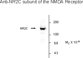
| NMDA NR2C Subunit antibody labels the 140k NR2C band in Western blots of cerebellum, and it labels the 180k NR2A and NR2B subunits of the NMDAR. Immunolabeling blocked by preadsorption of antibody with the N-terminal fusion protein of the NMDAR2C that was used to generate the antibody. Antibody dilutions and tissue load should be based on tissue type and expected expression level of the protein. Initial recommended range of dilutions: 1:500 to 1:2000. Applications include Dot Blots (DB), Immunohistochemistry (IHC), Immunoprecipitation (IP) and Western Blots (WB). Human, mouse, and rat have 100% amino acid sequence identity with the antigen used to raise the antibody. When internally tested under ideal conditions the working dilutions were 1:1000 for IHC, DB and WB.: |
|
| Species Reactivity | HumanRat |
| Concentration | 1mg/ml |
| Gene Symbol | Grin2c |
|---|---|
| Alternative Names | NR2C GluN2C N-methyl D-aspartate receptor subtype 2C |
Application
NMDA NR2C Subunit Antibody| Application Notes | NMDA NR2C Subunit antibody labels the 140k NR2C band in Western blots of cerebellum, and it labels the 180k NR2A and NR2B subunits of the NMDAR. Immunolabeling blocked by preadsorption of antibody with the N-terminal fusion protein of the NMDAR2C that was used to generate the antibody. Antibody dilutions and tissue load should be based on tissue type and expected expression level of the protein. Initial recommended range of dilutions: 1:500 to 1:2000. Applications include Dot Blots (DB), Immunohistochemistry (IHC), Immunoprecipitation (IP) and Western Blots (WB). Human, mouse, and rat have 100% amino acid sequence identity with the antigen used to raise the antibody. When internally tested under ideal conditions the working dilutions were 1:1000 for IHC, DB and WB.: |
|---|
| Form | Liquid |
|---|---|
| Storage Instructions | NMDA NR2C Subunit antibody can be stored at -20˚C. After reconstitution in 50 μl PBS, the antibody should be aliquot and stored at -20˚C and is stable at -20˚C for at least 1 year. Ambient shipment is suitable for both international and domestic. |
| Storage Buffer | phosphate buffered saline , pH 7.4, 150mM NaCl, 0.02% sodium azide and 50% glycerol. |


