Application
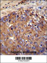
NOS2A Antibody immunohistochemistry analysis in formalin fixed and paraffin embedded human hepatocarcinoma followed by peroxidase conjugation of the secondary antibody and DAB staining.Application
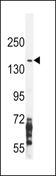
Western blot analysis in CEM cell line lysates (35ug/lane).Application
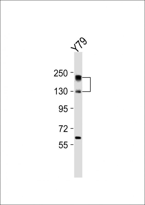
Western Blot at 1:2000 dilution + Y79 whole cell lysates Lysates/proteins at 20 ug per lane.Application
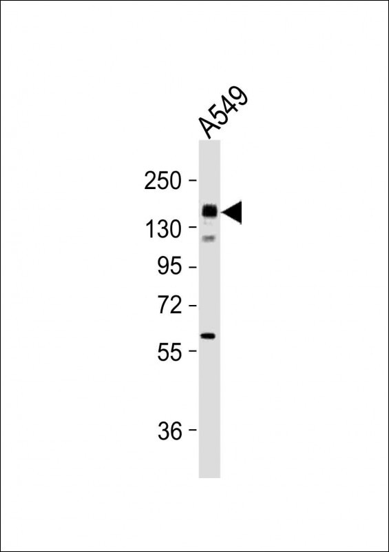
Western Blot at 1:2000 dilution + A549 whole cell lysates Lysates/proteins at 20 ug per lane.Application
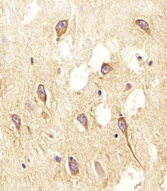
Antibody staining NOS2A in Human brain tissue sections by Immunohistochemistry (IHC-P - paraformaldehyde-fixed, paraffin-embedded sections).Application
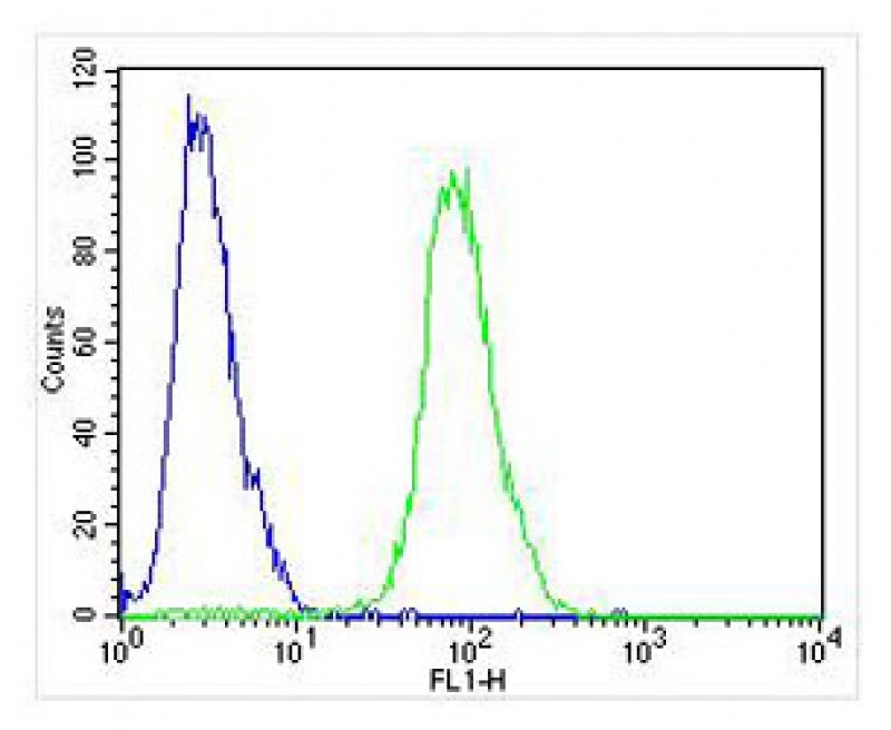
Overlay histogram showing Jurkat cells stained with Antibody (green line). The cells were fixed with 4% paraformaldehyde (10 min) and then permeabilized with 90% methanol for 10 min. The cells were then icubated in 2% bovine serum albumin to block non-specific protein-protein interactions followed by the antibody (1:25 dilution) for 60 min at 37ºC. The secondary antibody used was Alexa Fluor 488 gApplication
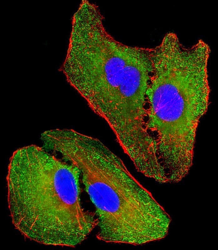
Immunofluorescent analysis of 4% paraformaldehyde-fixed, 0. 1% Triton X-100 permeabilized A549 (human lung adenocarcinoma epithelial cell line) cells labeling Pdx1 with antibody at 1/25 dilution, followed by Dylight 488-conjugated goat anti-rabbit IgG (NK179883) secondary antibody at 1/200 dilution (green). Immunofluorescence image showing cytoplasm staining on A549 cell line. Cytoplasmic actin is| Product Name | NOS2A Antibody |
|---|---|
| Antibody Type | Primary Antibodies |
| Antigen Alias | Nitric oxide synthase, inducible, Hepatocyte NOS, HEP-NOS, Inducible NO synthase, Inducible NOS, iNOS, NOS type II, Peptidyl-cysteine S-nitrosylase NOS2, NOS2, NOS2A |
| Product description | Nitric oxide is a reactive free radical which acts as a biologic mediator in several processes, including neurotransmission and antimicrobial and antitumoral activities. This gene encodes a nitric oxide synthase which is expressed in liver and is inducible by a combination of lipopolysaccharide and certain cytokines. Three related pseudogenes are located within the Smith-Magenis syndrome region on chromosome 17.1) Ryk, C., et al. J. Urol. 184(5):2150-2157(2010) |
| Immunogen | This NOS2A antibody is generated from rabbits immunized with a KLH conjugated synthetic peptide between 830-860 amino acids from the Central region of human NOS2A. |
| Clonality | Polyclonal |
|---|---|
| Isotype | Ig |
| Host Species | Rabbit |
| Tested Applications | FACSIFIHC-PWB |
| For IF starting dilution is: 1:25 For FACS starting dilution is: 1:25 For IHC-P starting dilution is: 1:25 For WB starting dilution is: 1:1000: |
|
| Species Reactivity | Human |
| Concentration | 1mg/ml |
| Purification | Affinity purified |
| Gene Symbol | NOS2 |
|---|---|
| Alternative Names | Nitric oxide synthase inducible Hepatocyte NOS HEP-NOS Inducible NO synthase Inducible NOS iNOS NOS type II Peptidyl-cysteine S-nitrosylase NOS2 NOS2 NOS2A |
| Molecular Weight(MW) | 131 kDa |
Application
NOS2A Antibody immunohistochemistry analysis in formalin fixed and paraffin embedded human hepatocarcinoma followed by peroxidase conjugation of the secondary antibody and DAB staining.
Application
Western blot analysis in CEM cell line lysates (35ug/lane).
Application
Western Blot at 1:2000 dilution + Y79 whole cell lysates Lysates/proteins at 20 ug per lane.
Application
Western Blot at 1:2000 dilution + A549 whole cell lysates Lysates/proteins at 20 ug per lane.
Application
Antibody staining NOS2A in Human brain tissue sections by Immunohistochemistry (IHC-P - paraformaldehyde-fixed, paraffin-embedded sections).
Application
Overlay histogram showing Jurkat cells stained with Antibody (green line). The cells were fixed with 4% paraformaldehyde (10 min) and then permeabilized with 90% methanol for 10 min. The cells were then icubated in 2% bovine serum albumin to block non-specific protein-protein interactions followed by the antibody (1:25 dilution) for 60 min at 37ºC. The secondary antibody used was Alexa Fluor 488 g
Application
Immunofluorescent analysis of 4% paraformaldehyde-fixed, 0. 1% Triton X-100 permeabilized A549 (human lung adenocarcinoma epithelial cell line) cells labeling Pdx1 with antibody at 1/25 dilution, followed by Dylight 488-conjugated goat anti-rabbit IgG (NK179883) secondary antibody at 1/200 dilution (green). Immunofluorescence image showing cytoplasm staining on A549 cell line. Cytoplasmic actin is| Application Notes | For IF starting dilution is: 1:25 For FACS starting dilution is: 1:25 For IHC-P starting dilution is: 1:25 For WB starting dilution is: 1:1000: |
|---|
| Form | Liquid |
|---|---|
| Storage Instructions | Store at 4˚C for three months and -20˚C, stable for up to one year. As with all antibodies care should be taken to avoid repeated freeze thaw cycles. Antibodies should not be exposed to prolonged high temperatures. |
| Storage Buffer | Supplied in PBS with 0.09% (W/V) sodium azide. |


 Data sheet for OM286393