Application
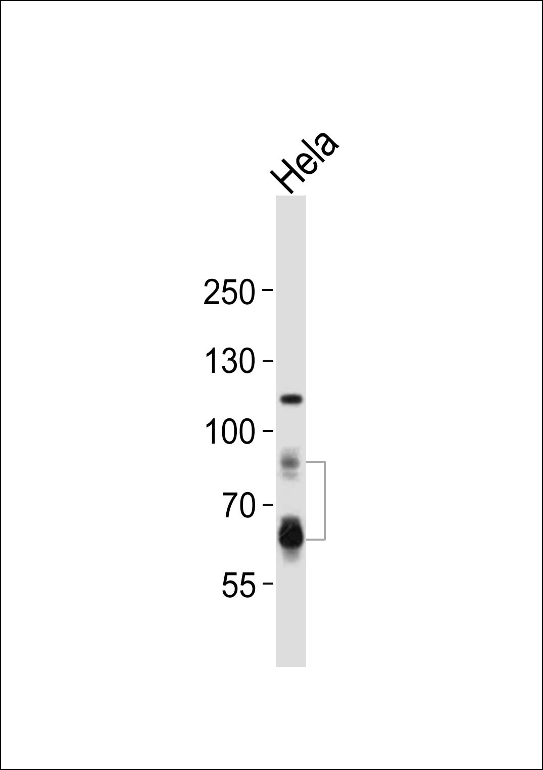
Western blot analysis of lysate from Hela cell line, using PCSK9 Antibody at 1:1000.Application
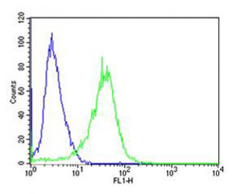
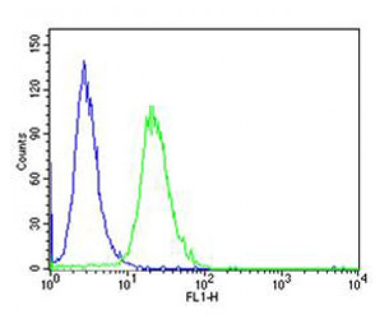
Flow cytometric analysis of A431 cells using PCSK9 Antibody (green) compared to an isotype control of rabbit IgG(blue). Antibody was diluted at 1:25 dilution. An Alexa Fluor 488 goat anti-rabbit lgG at 1:400 dilution was used as the secondary antibody.Application
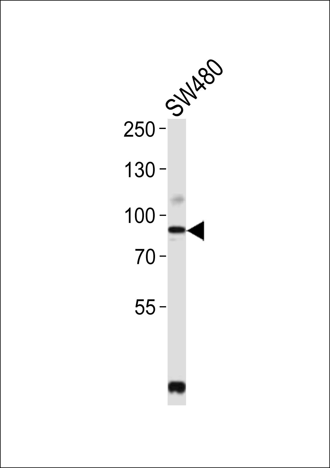
Western blot analysis of lysate from SW480 cell line, using PCSK9 Antibody at 1:2000.Application
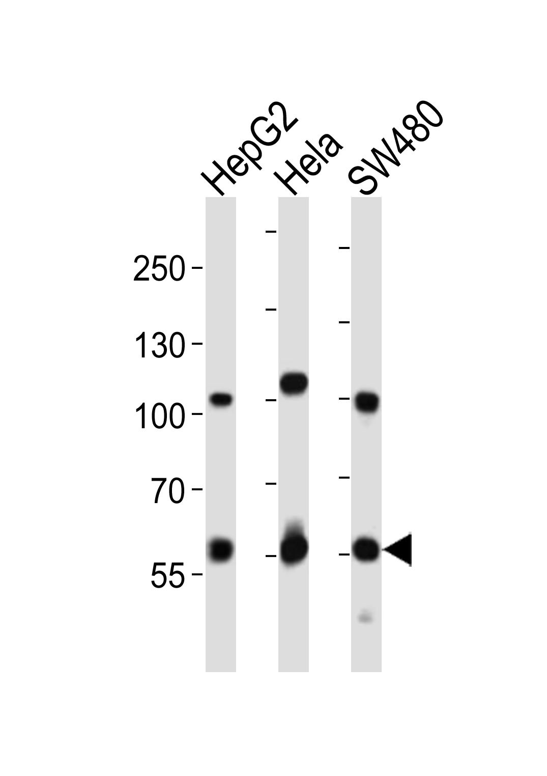
Western blot analysis of lysates from HepG2, Hela, SW480 cell line (from left to right), using PCSK9 Antibody at 1:1000 at each lane.Application
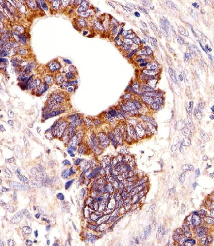
Immunohistochemical analysis of paraffin-embedded H. colorectal carcinoma section using PCSK9 Antibody . Antibody was diluted at 1:25 dilution. A undiluted biotinylated goat polyvalent antibody was used as the secondary, followed by DAB staining.Application
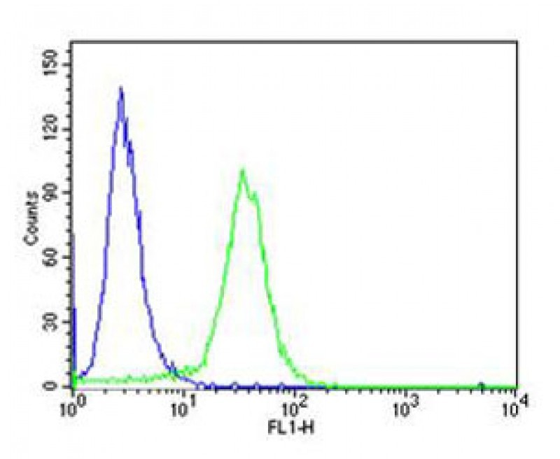
Flow cytometric analysis of HeLa cells using PCSK9 Antibody (green) compared to an isotype control of rabbit IgG(blue). Antibody was diluted at 1:25 dilution. An Alexa Fluor 488 goat anti-rabbit lgG at 1:400 dilution was used as the secondary antibody.Application
Flow cytometric analysis of HeLa cells using PCSK9 Antibody (green) compared to an isotype control of rabbit IgG(blue). Antibody was diluted at 1:25 dilution. An Alexa Fluor 488 goat anti-rabbit lgG at 1:400 dilution was used as the secondary antibody.| Product Name | PCSK9 Antibody |
|---|---|
| Antibody Type | Primary Antibodies |
| Antigen Alias | Proprotein convertase subtilisin/kexin type 9, 3421-, Neural apoptosis-regulated convertase 1, NARC-1, Proprotein convertase 9, PC9, Subtilisin/kexin-like protease PC9, PCSK9, NARC1 |
| Product description | PCSK9 is a proprotein convertase belonging to the proteinase K subfamily of the secretory subtilase family. This protein is synthesized as a soluble zymogen that undergoes autocatalytic intramolecular processing in the endoplasmic reticulum. The protein may function as a proprotein convertase. The protein plays a role in cholesterol homeostasis and may have a role in the differentiation of cortical neurons.1) Abifadel,M., Rabes,J.P. Hum. Mutat. 30 (7), E682-E691 (2009) |
| Immunogen | This PCSK9 antibody is generated from rabbits immunized with a KLH conjugated synthetic peptide between 144-173 amino acids from the N-terminal region of human PCSK9. |
| Clonality | Polyclonal |
|---|---|
| Isotype | Ig |
| Host Species | Rabbit |
| Tested Applications | FACSIHC-PWB |
| For FACS starting dilution is: 1:25 For IHC-P starting dilution is: 1:25 For WB starting dilution is: 1:2000: |
|
| Species Reactivity | Human |
| Concentration | 1mg/ml |
| Purification | Affinity purified |
| Gene Symbol | PCSK9 |
|---|---|
| Alternative Names | Proprotein convertase subtilisin/kexin type 9 3421- Neural apoptosis-regulated convertase 1 NARC-1 Proprotein convertase 9 PC9 Subtilisin/kexin-like protease PC9 PCSK9 NARC1 |
| Molecular Weight(MW) | 74 kDa |
Application
Western blot analysis of lysate from Hela cell line, using PCSK9 Antibody at 1:1000.
Application
Flow cytometric analysis of A431 cells using PCSK9 Antibody (green) compared to an isotype control of rabbit IgG(blue). Antibody was diluted at 1:25 dilution. An Alexa Fluor 488 goat anti-rabbit lgG at 1:400 dilution was used as the secondary antibody.
Application
Western blot analysis of lysate from SW480 cell line, using PCSK9 Antibody at 1:2000.
Application
Western blot analysis of lysates from HepG2, Hela, SW480 cell line (from left to right), using PCSK9 Antibody at 1:1000 at each lane.
Application
Immunohistochemical analysis of paraffin-embedded H. colorectal carcinoma section using PCSK9 Antibody . Antibody was diluted at 1:25 dilution. A undiluted biotinylated goat polyvalent antibody was used as the secondary, followed by DAB staining.
Application
Flow cytometric analysis of HeLa cells using PCSK9 Antibody (green) compared to an isotype control of rabbit IgG(blue). Antibody was diluted at 1:25 dilution. An Alexa Fluor 488 goat anti-rabbit lgG at 1:400 dilution was used as the secondary antibody.
Application
Flow cytometric analysis of HeLa cells using PCSK9 Antibody (green) compared to an isotype control of rabbit IgG(blue). Antibody was diluted at 1:25 dilution. An Alexa Fluor 488 goat anti-rabbit lgG at 1:400 dilution was used as the secondary antibody.| Application Notes | For FACS starting dilution is: 1:25 For IHC-P starting dilution is: 1:25 For WB starting dilution is: 1:2000: |
|---|
| Form | Liquid |
|---|---|
| Storage Instructions | Store at 4˚C for three months and -20˚C, stable for up to one year. As with all antibodies care should be taken to avoid repeated freeze thaw cycles. Antibodies should not be exposed to prolonged high temperatures. |
| Storage Buffer | Supplied in PBS with 0.09% (W/V) sodium azide. |


 Data sheet for OM287601