

Application
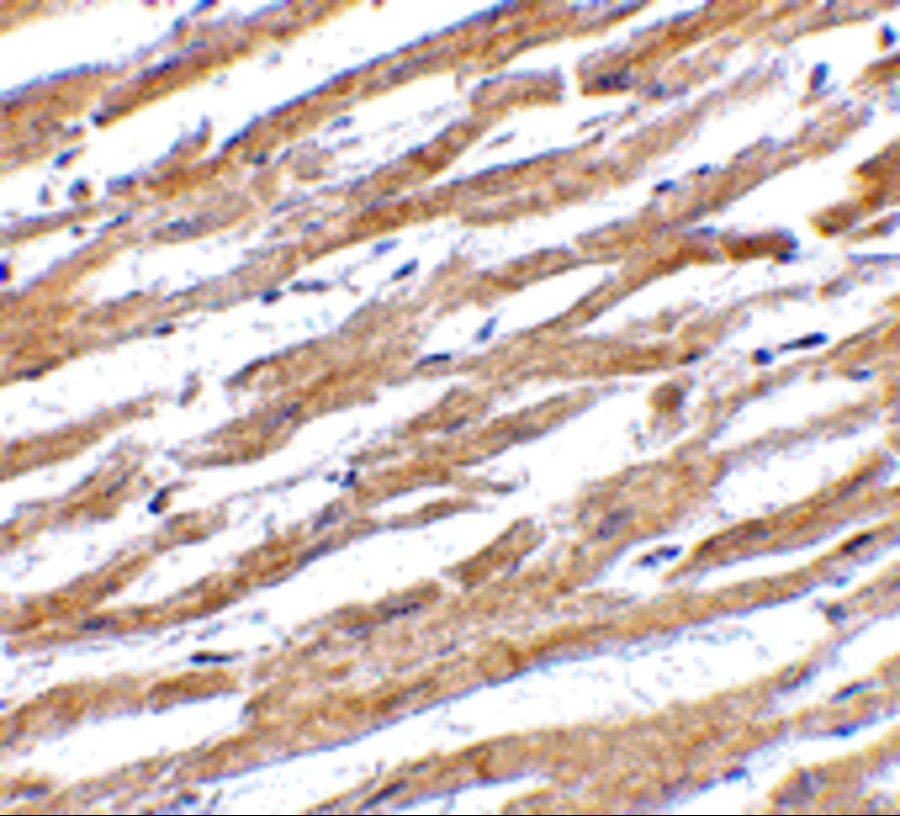
Immunohistochemistry of PD-L1 in human heart tissue with PD-L1 antibodyApplication
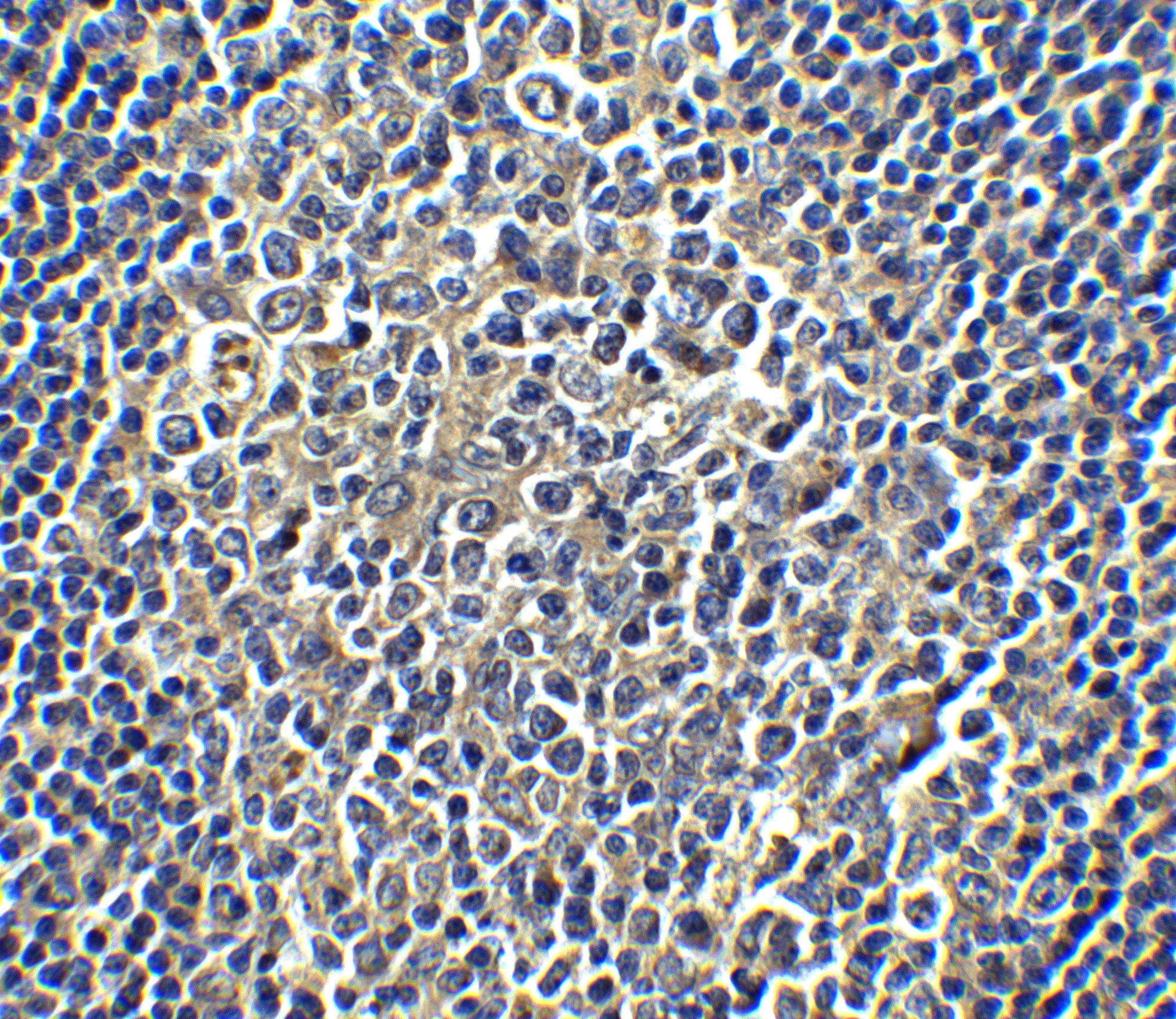
Immunohistochemistry of PD-L1 in human tonsil tissue with PD-L1 antibodyApplication
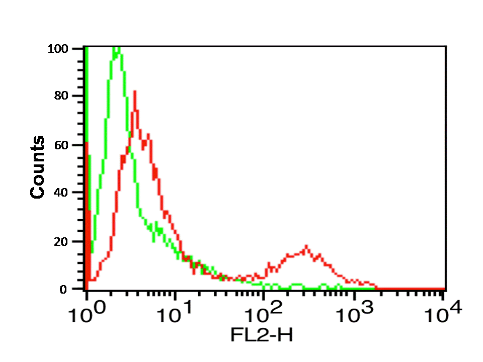
Flow cytometry analysis of A-20 cells using PD-L1 antibodyl. Green: Isotype control. Red : PD-L1 antibody.Application
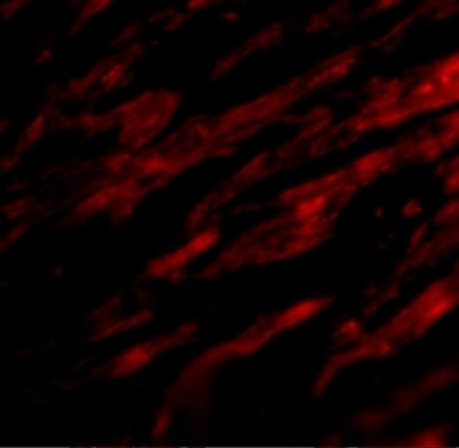
Immunofluorescence of PD-L1 in Human Heart cells with PD-L1 antibodyWB
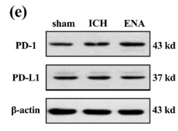
(e) The representative western blot bands of PD-1 and PD-L1 around lesion area.IF
(h and i) PD1 and PD-L1 cells around the lesion area were labeled by immunofluorescence (n = 3, each sample was chosen two fields of view).IF
(h and i) PD1 and PD-L1 cells around the lesion area were labeled by immunofluorescence (n = 3, each sample was chosen two fields of view).| Product Name | PD-L1 Antibody |
|---|---|
| Antibody Type | Primary Antibodies |
| Product description | PD-L1 Antibody: Cell-mediated immune responses are initiated by T lymphocytes that are themselves stimulated by cognate peptides bound to MHC molecules on antigen-presenting cells (APC). T-cell activation is generally self-limited as activated T cells express receptors such as PD-1 (also known as PDCD-1) that mediate inhibitory signals from the APC. PD-1 can bind two different but related ligands, PD-L1 and PD-L2. PD-L1 is a B7-related protein that inhibits cell-mediated immune responses by reducing the secretion of IL-2 and IL-10 from memory T cells. This suggests that PD-L1 may be useful in reducing allogenic CD4+ memory T-cell responses to endothelial cells, thereby reducing the likelihood of host immune responses to allografts. At least two isoforms of PD-L1 are known to exist; this antibody is specific to the larger isoform.1) Holling TM, Schooten E, and van Den Elsing PJ. Function and regulation of MHC class II molecules in T-lymphocytes: of mice and men. Hum. Immunol. 2004; 65:282-90. |
| Immunogen | PD-L1 antibody was raised against a 17 amino acid synthetic peptide from near the center of human PD-L1. The immunogen is located within amino acids 60 - 110 of PD-L1. |
| Modification | Acetylated |
| Clonality | Polyclonal |
|---|---|
| Isotype | IgG |
| Host Species | Rabbit |
| Tested Applications | ELISAFACSIFIHC-PWB |
| WB:1:500~1:2000 IF:1:100~1:500 IHC-p:1:50~1:200 |
|
| Species Reactivity | HumanMouseRat |
| Concentration | 1mg/ml |
| Purification | Affinity purified |
| Gene Symbol | CD274 |
|---|---|
| Alternative Names | PD-L1 Antibody: B7-H B7H1 PDL1 PD-L1 PDCD1L1 PDCD1LG1 Programmed cell death 1 ligand 1 B7 homolog 1 |
| Molecular Weight(MW) | Predicted: 32 kDa Observed: 37 kDa |
| Sequence Similarities | 3) Cat. No. 10-501 - Human Heart Tissue Slide |
| Tissue Specificity | PD-L1 antibody has no cross-reactivity to PD-L2. |
Application
Immunohistochemistry of PD-L1 in human heart tissue with PD-L1 antibody
Application
Immunohistochemistry of PD-L1 in human tonsil tissue with PD-L1 antibody
Application
Flow cytometry analysis of A-20 cells using PD-L1 antibodyl. Green: Isotype control. Red : PD-L1 antibody.
Application
Immunofluorescence of PD-L1 in Human Heart cells with PD-L1 antibody
WB
(e) The representative western blot bands of PD-1 and PD-L1 around lesion area.
IF
(h and i) PD1 and PD-L1 cells around the lesion area were labeled by immunofluorescence (n = 3, each sample was chosen two fields of view).
IF
(h and i) PD1 and PD-L1 cells around the lesion area were labeled by immunofluorescence (n = 3, each sample was chosen two fields of view).| Positive Control | 1) Cat. No. 1207 - Raji Cell Lysate |
|---|---|
| Application Notes | WB:1:500~1:2000 IF:1:100~1:500 IHC-p:1:50~1:200 |
| Form | Liquid |
|---|---|
| Storage Instructions | PD-L1 antibody can be stored at 4˚C for three months and -20˚C, stable for up to one year. As with all antibodies care should be taken to avoid repeated freeze thaw cycles. Antibodies should not be exposed to prolonged high temperatures. |
| Storage Buffer | PD-L1 Antibody is supplied in PBS containing 0.02% sodium azide. |


