Application
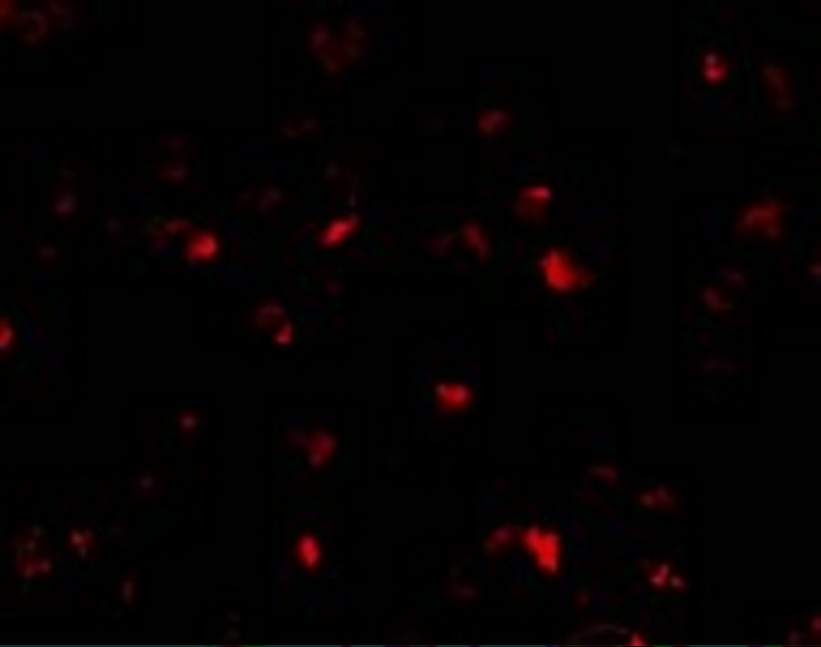
Immunofluorescence of PHAP I in Raji cells with PHAP I antibody at 10 μg/mL.Application
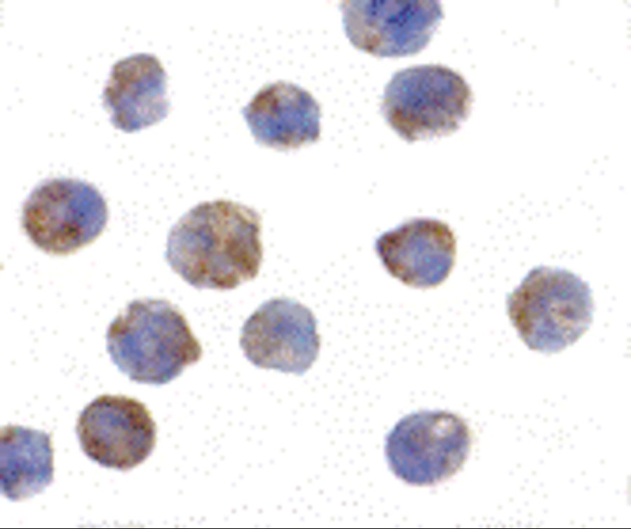
Immunocytochemistry of PHAP I in Raji cells with PHAP I antibody at 2 μg/mL.Application
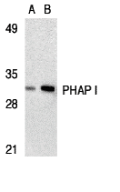
Western blot analysis of PHAP I expression in human Raji cell lysate with PHAP antibody I at 2 μg/mL (lane A) and 4 μg/mL (lane B), respectively.| Product Name | PHAP I Antibody |
|---|---|
| Antibody Type | Primary Antibodies |
| Antigen Alias | PHAP I Antibody: LANP, MAPM, PP32, HPPCn, PHAP1, PHAPI, I1PP2A, C15orf1, LANP, Acidic leucine-rich nuclear phosphoprotein 32 family member A, Acidic nuclear phosphoprotein pp32 |
| Product description | PHAP I Antibody: Apoptosis is related to many diseases and development. Caspase-9 plays a central role in cell death induced by a variety of apoptosis activators. Cytochrome c, after released from mitochondria, binds to Apaf-1, which forms an apoptosome that in turn binds to and activate procaspase-9. Activated caspase-9 cleaves and activates the effector caspases (caspase-3, -6 and -7), which are responsible for the proteolytic cleavage of many key proteins in apoptosis. The tumor suppressor putative HLA-DR-associated proteins (PHAPs) were recently identified as important regulators of mitochondrion apoptosis. PHAP appears to facilitate apoptosome-medicated caspase-9 activation and to stimulate the mitochondrial apoptotic pathway. PHAP was also shown to oppose both Ras- and Myc-medicated cell transformation.1) Jiang X, Kim HE, Shu H, Zhao Y, Zhang H, Kofron J, Donnelly J, Burns D, Ng SC , Rosenberg S, Wang X. Distinctive roles of PHAP proteins and prothymosin-α in a death regulatory pathway. Science. 2003;299(5604):223-6. |
| Immunogen | PHAP I antibody was raised with a synthetic peptide corresponding to amino acids close to carboxy terminus of human PHAP I. The sequence is identical between human and rat PHAP I. The immunogen is located within the last 50 amino acids of PHAP I. |
| Clonality | Polyclonal |
|---|---|
| Isotype | IgG |
| Host Species | Rabbit |
| Tested Applications | ELISAICCIFWB |
| PHAP I antibody can be used for detection of PHAP I by Western blot at 1 - 2 μg/mL. A band at approximately 32 kDa can be detected. Antibody can also be used for immunocytochemistry starting at 2 μg/mL. For immunofluorescence start at 10 μg/mL.: |
|
| Species Reactivity | HumanMouse |
| Concentration | 1mg/ml |
| Purification | Affinity purified |
| Gene Symbol | ANP32A |
|---|---|
| Alternative Names | PHAP I Antibody: LANP MAPM PP32 HPPCn PHAP1 PHAPI I1PP2A C15orf1 LANP Acidic leucine-rich nuclear phosphoprotein 32 family member A Acidic nuclear phosphoprotein pp32 |
| Molecular Weight(MW) | 32 kDa |
| Sequence Similarities | 2) Cat. No. 17-007 - Raji Cell Slide |
| Tissue Specificity | This polyclonal antibody has no cross-reaction to PHAP I2a and PHAP III. |
Application
Immunofluorescence of PHAP I in Raji cells with PHAP I antibody at 10 μg/mL.
Application
Immunocytochemistry of PHAP I in Raji cells with PHAP I antibody at 2 μg/mL.
Application
Western blot analysis of PHAP I expression in human Raji cell lysate with PHAP antibody I at 2 μg/mL (lane A) and 4 μg/mL (lane B), respectively.| Positive Control | 1) Cat. No. 1207 - Raji Cell Lysate |
|---|---|
| Application Notes | PHAP I antibody can be used for detection of PHAP I by Western blot at 1 - 2 μg/mL. A band at approximately 32 kDa can be detected. Antibody can also be used for immunocytochemistry starting at 2 μg/mL. For immunofluorescence start at 10 μg/mL.: |
| Form | Liquid |
|---|---|
| Storage Instructions | PHAP I antibody can be stored at 4˚C for three months and -20˚C, stable for up to one year. As with all antibodies care should be taken to avoid repeated freeze thaw cycles. Antibodies should not be exposed to prolonged high temperatures. |
| Storage Buffer | PHAP I Antibody is supplied in PBS containing 0.02% sodium azide. |


