Application
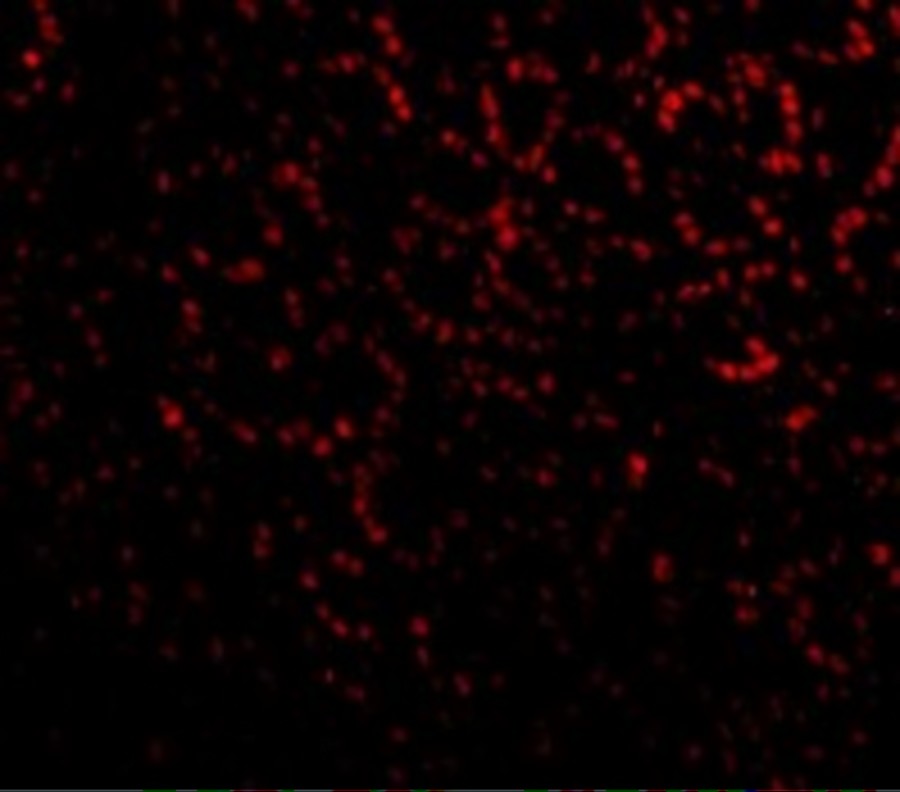
Immunofluorescence of PHAP I in Mouse Small Intestine cells with PHAP I antibody at 20 μg/mL.Application
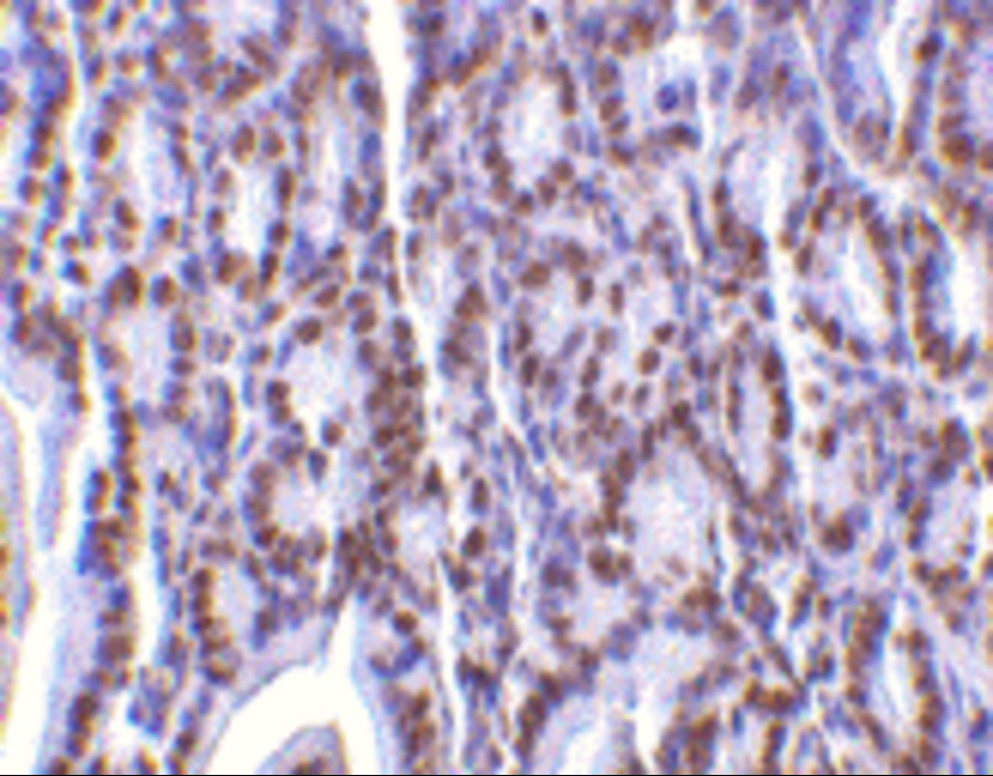
Immunohistochemistry of PHAP I in mouse small intestine tissue with PHAP I antibody at 2 μg/mL.Application
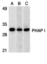
Western blot analysis of PHAP I expression in human Raji cell (A), mouse (B) and rat (C) testis tissue llysates with PHAP I antibody at 1 μg/mL.| Product Name | PHAP I Antibody |
|---|---|
| Antibody Type | Primary Antibodies |
| Antigen Alias | PHAP I Antibody: LANP, MAPM, PP32, HPPCn, PHAP1, PHAPI, I1PP2A, C15orf1, LANP, Acidic leucine-rich nuclear phosphoprotein 32 family member A, Acidic nuclear phosphoprotein pp32 |
| Product description | PHAP I Antibody: Apoptosis is related to many diseases and development. Caspase-9 plays a central role in cell death induced by a variety of apoptosis activators. Cytochrome c, after released from mitochondria, binds to Apaf-1, which forms an apoptosome that in turn binds to and activate procaspase-9. Activated caspase-9 cleaves and activates the effector caspases (caspase-3, -6 and -7), which are responsible for the proteolytic cleavage of many key proteins in apoptosis. The tumor suppressor putative HLA-DR-associated proteins (PHAPs) were recently identified as important regulators of mitochondrion apoptosis. PHAP appears to facilitate apoptosome-medicated caspase-9 activation and to stimulate the mitochondrial apoptotic pathway. PHAP was also shown to oppose both Ras- and Myc-medicated cell transformation.1) Jiang X, Kim HE, Shu H, Zhao Y, Zhang H, Kofron J, Donnelly J, Burns D, Ng SC , Rosenberg S, Wang X. Distinctive roles of PHAP proteins and prothymosin-α in a death regulatory pathway. Science. 2003;299(5604):223-6. |
| Immunogen | PHAP I antibody was raised with a synthetic peptide corresponding to amino acids at carboxy terminus of human PHAP I. The immunogen is located within the last 50 amino acids of PHAP I. |
| Clonality | Polyclonal |
|---|---|
| Isotype | IgG |
| Host Species | Rabbit |
| Tested Applications | ELISAIFIHC-PWB |
| PHAP I antibody can be used for detection of PHAP I by Western blot at 1 μg/mL. A band at approximately 32 kDa can be detected. Antibody can also be used for immunohistochemistry starting at 2 μg/mL. For immunofluorescence start at 2 μg/mL.: |
|
| Species Reactivity | HumanMouseRat |
| Concentration | 1mg/ml |
| Purification | Affinity purified |
| Gene Symbol | ANP32A |
|---|---|
| Alternative Names | PHAP I Antibody: LANP MAPM PP32 HPPCn PHAP1 PHAPI I1PP2A C15orf1 LANP Acidic leucine-rich nuclear phosphoprotein 32 family member A Acidic nuclear phosphoprotein pp32 |
| Molecular Weight(MW) | 32 kDa |
| Sequence Similarities | 4) Cat. No. 1408 - Mouse Small Intestine Tissue Lysate |
| Tissue Specificity | This polyclonal antibody has no cross-reaction to PHAP I2a and PHAP III. |
Application
Immunofluorescence of PHAP I in Mouse Small Intestine cells with PHAP I antibody at 20 μg/mL.
Application
Immunohistochemistry of PHAP I in mouse small intestine tissue with PHAP I antibody at 2 μg/mL.
Application
Western blot analysis of PHAP I expression in human Raji cell (A), mouse (B) and rat (C) testis tissue llysates with PHAP I antibody at 1 μg/mL.| Positive Control | 1) Cat. No. 1207 - Raji Cell Lysate |
|---|---|
| Application Notes | PHAP I antibody can be used for detection of PHAP I by Western blot at 1 μg/mL. A band at approximately 32 kDa can be detected. Antibody can also be used for immunohistochemistry starting at 2 μg/mL. For immunofluorescence start at 2 μg/mL.: |
| Form | Liquid |
|---|---|
| Storage Instructions | PHAP I antibody can be stored at 4˚C for three months and -20˚C, stable for up to one year. As with all antibodies care should be taken to avoid repeated freeze thaw cycles. Antibodies should not be exposed to prolonged high temperatures. |
| Storage Buffer | PHAP I Antibody is supplied in PBS containing 0.02% sodium azide. |


