Application
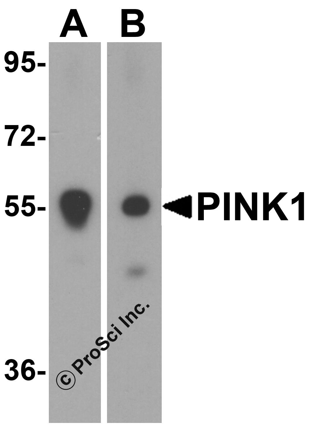
Western blot analysis of PINK1 in (A) mouse and (B) rat spleen tissue lysate with PINK1 antibody at 1 μg/mL.Application
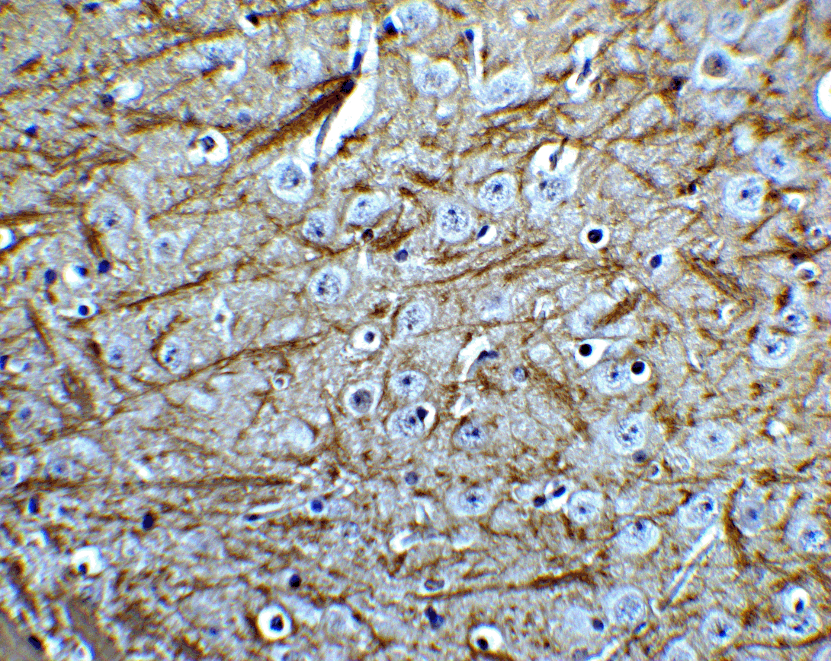
Immunohistochemistry of PINK1 in mouse brain tissue with PINK1 Antibodyat 5 μg/mL.Application
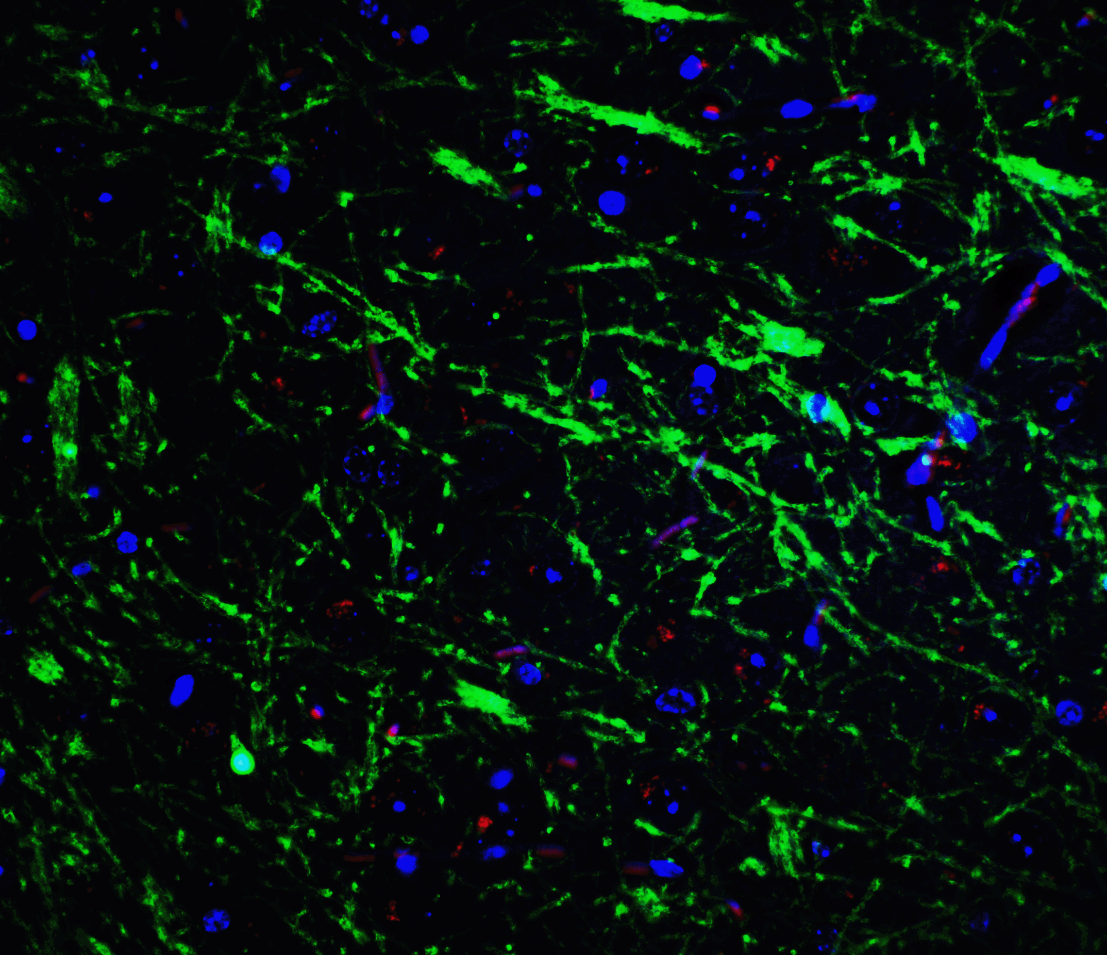
Immunofluorescence of PINK1 in mouse brain tissue with PINK1 Antibodyat 20 μg/mL.Application
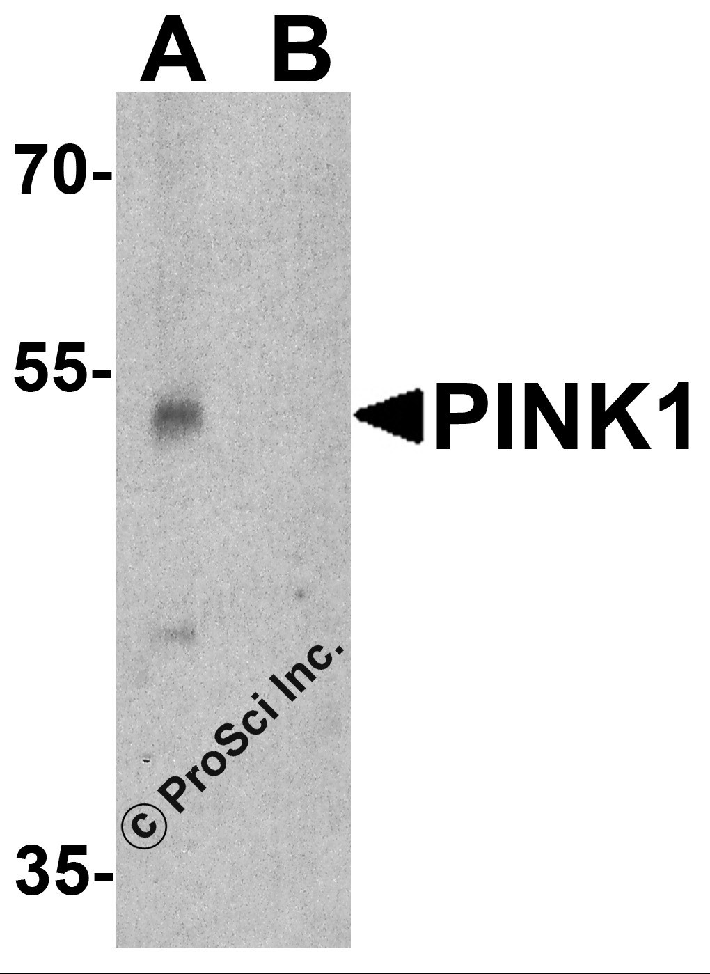
Western blot analysis of PINK1 in A431 cell lysate with PINK1 antibody at 1 μg/ml in (A) the absence and (B) the presence of blocking peptide.| Product Name | PINK1 Antibody |
|---|---|
| Antibody Type | Primary Antibodies |
| Antigen Alias | PINK1 Antibody: BRPK, PARK6BRPK |
| Product description | The PTEN-induced putative kinase 1 (PINK1) is a serine/threonine protein kinase that localizes to mitochondria and is thought to protect cells from stress-induced mitochondrial dysfunction (reviewed in 1). PINK1 recruits the E3 ubiquitin ligase Parkin to mitochondria to initiate mitophagy, an autophagic process that clears damaged mitochondria within a cell (2). PINK1 is cleaved by the mitochondrial protease PARL (3). Mutations in this gene cause one form of autosomal recessive early-onset Parkinson disease (4).1) Matsuda S, Kitagishi Y, and Kobayashi M. Function and characteristics of PINK1 in mitochondria. Oxid. Med. Cell. Longev. 2013;601587. |
| Immunogen | PINK1 antibody was raised against a 16 amino acid peptide near the amino terminus of human PINK1. The immunogen is located within amino acids 120 - 170 of PINK1. |
| Clonality | Polyclonal |
|---|---|
| Isotype | IgG |
| Host Species | Rabbit |
| Tested Applications | ELISAIFIHC-PWB |
| WB:1:500~1:1000 IHC-p:1:50~1:200 IF:1:50~1:500 |
|
| Species Reactivity | HumanMouseRat |
| Concentration | 1mg/ml |
| Purification | Affinity purified |
| Gene Symbol | PINK1 |
|---|---|
| Alternative Names | PINK1 Antibody: BRPK PARK6BRPK |
| Molecular Weight(MW) | Predicted: 55 kDa Observed: 53 kDa |
| Sequence Similarities | 3) Cat. No. 1466 - Rat Spleen Tissue Lysate |
| Tissue Specificity | PINK1 antibody is human, mouse and rat reactive. At least two isoforms are known to exist; this antibody will only detect the longer isoform. PINK1 antibody will detect the cleaved and uncleaved form of PINK1. |
Application
Western blot analysis of PINK1 in (A) mouse and (B) rat spleen tissue lysate with PINK1 antibody at 1 μg/mL.
Application
Immunohistochemistry of PINK1 in mouse brain tissue with PINK1 Antibodyat 5 μg/mL.
Application
Immunofluorescence of PINK1 in mouse brain tissue with PINK1 Antibodyat 20 μg/mL.
Application
Western blot analysis of PINK1 in A431 cell lysate with PINK1 antibody at 1 μg/ml in (A) the absence and (B) the presence of blocking peptide.| Positive Control | 1) Cat. No. 1202 - A431 Cell Lysate |
|---|---|
| Application Notes | WB:1:500~1:1000 IHC-p:1:50~1:200 IF:1:50~1:500 |
| Form | Liquid |
|---|---|
| Storage Instructions | PINK1 antibody can be stored at 4˚C for three months and -20˚C, stable for up to one year. |
| Storage Buffer | PINK1 antibody is supplied in PBS containing 0.02% sodium azide. |


