Application
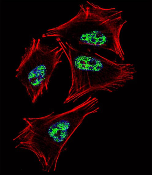
Fluorescent confocal image of A2058 cell stained with PROX1 Antibody .A2058 cells were fixed with 4% PFA (20 min), permeabilized with Triton X-100 (0.1%, 10 min), then incubated with PROX1 primary antibody (1:25). For secondary antibody, Alexa Fluor 488 conjugated donkey anti-rabbit antibody (green) was used (1:400).Cytoplasmic actin was counterstained with Alexa Fluor 555 (red) conjugated PhalloiApplication
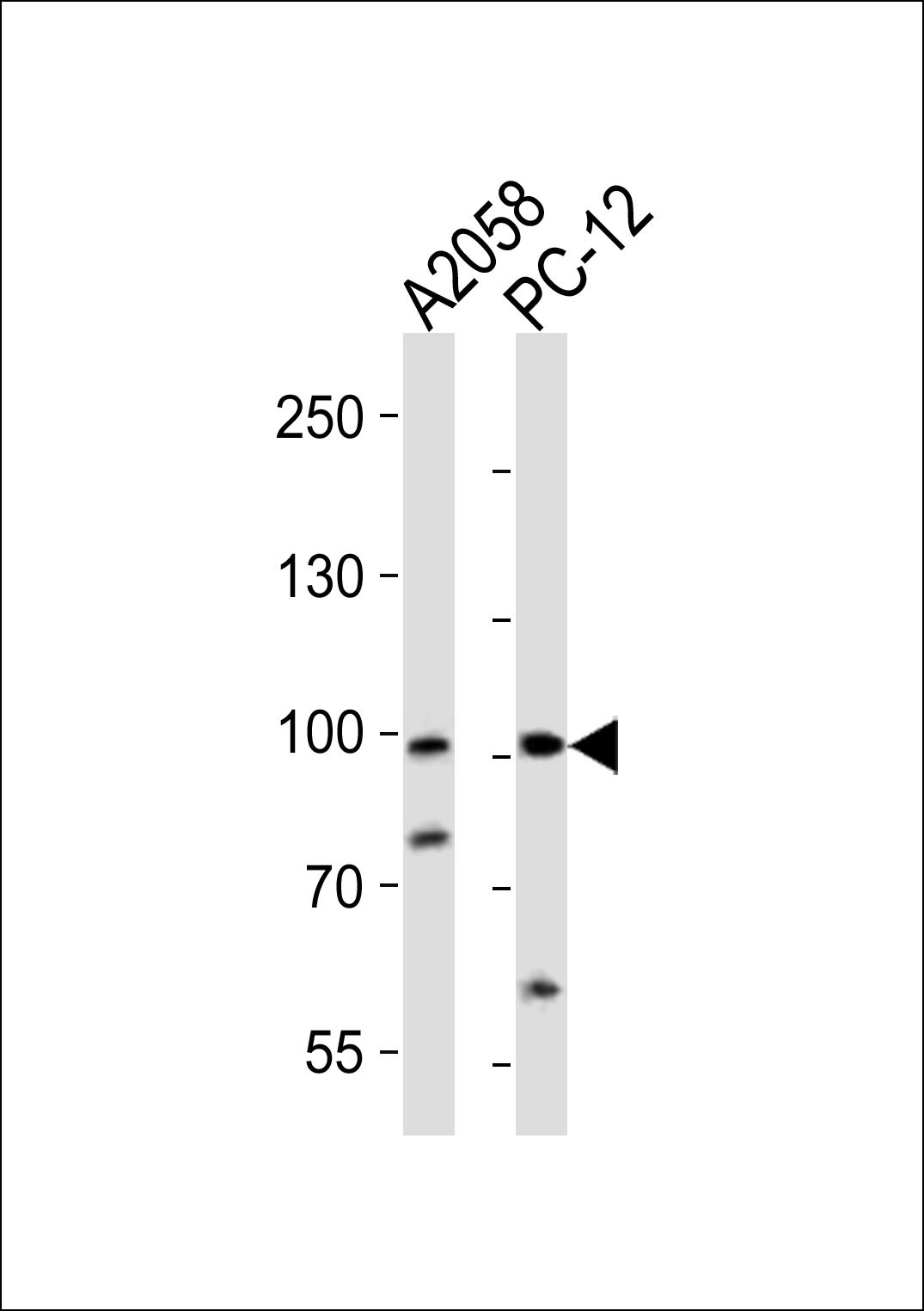
Western blot analysis of lysates from A2058, rat PC-12 cell line (from left to right), using PROX1 Antibody at 1:1000 at each lane.Application
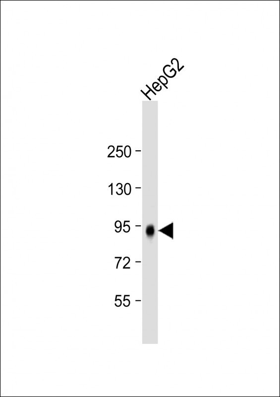
Western Blot at 1:2000 dilution + HepG2 whole cell lysate Lysates/proteins at 20 ug per lane.| Product Name | PROX1 Antibody |
|---|---|
| Antibody Type | Primary Antibodies |
| Antigen Alias | Prospero homeobox protein 1, Homeobox prospero-like protein PROX1, PROX-1, PROX1 |
| Product description | The expression pattern of the Prox1 homeo box gene suggests that it has a role in a variety of embryonic tissues, including lens. Analysis of mRNA reveals that Prox mRNA is present in many different human tissues and that lens demonstrated the highest level. Homozygous Prox1-null mice die at midgestation from multiple developmental defects, and a targeted effect on lens development has been reported. Prox1 inactivation caused abnormal cellular proliferation, downregulated expression of the cell cycle inhibitors Cdkn1b and Cdkn1c, misexpression of E-cadherin, and excessive apoptosis. Consequently, mutant lens cells failed to polarize and elongate properly, resulting in a hollow lens. The Prox1 gene is expressed in a subpopulation of endothelial cells that by budding and sprouting give rise to the lymphatic system. Prox1 appears to be a specific and required regulator of the development of the lymphatic system. Prox1 also has been document to be required for hepatocyte migration in the mouse. Loss of Prox1 results in a smaller liver with a reduced population of clustered hepatocytes. The homeodomain protein Prox1 regulates the egress of progenitor cells from the cell cycle in the embryonic mouse retina. Cells lacking Prox1 are less likely to stop dividing, and ectopic expression of Prox1 forces progenitor cells to exit the cell cycle. Prox1 acts as a key participant in progenitor-cell proliferation and cell-fate determination in the vertebrate retina.1) Nagai, H., et al., Genes Chromosomes Cancer 38(1):13-21 (2003). |
| Immunogen | This PROX1 antibody is generated from rabbits immunized with a KLH conjugated synthetic peptide between 492-522 amino acids from the C-terminal region of human PROX1. |
| Clonality | Polyclonal |
|---|---|
| Isotype | Ig |
| Host Species | Rabbit |
| Tested Applications | IFWB |
| For WB starting dilution is: 1:500 For IF starting dilution is: 1:10~50: |
|
| Species Reactivity | Human |
| Concentration | 1mg/ml |
| Gene Symbol | PROX1 |
|---|---|
| Alternative Names | Prospero homeobox protein 1 Homeobox prospero-like protein PROX1 PROX-1 PROX1 |
| Molecular Weight(MW) | 83 kDa |
| Sequence Similarities | Predicted species reactivity based on immunogen sequence: Mouse |
Application
Fluorescent confocal image of A2058 cell stained with PROX1 Antibody .A2058 cells were fixed with 4% PFA (20 min), permeabilized with Triton X-100 (0.1%, 10 min), then incubated with PROX1 primary antibody (1:25). For secondary antibody, Alexa Fluor 488 conjugated donkey anti-rabbit antibody (green) was used (1:400).Cytoplasmic actin was counterstained with Alexa Fluor 555 (red) conjugated Phalloi
Application
Western blot analysis of lysates from A2058, rat PC-12 cell line (from left to right), using PROX1 Antibody at 1:1000 at each lane.
Application
Western Blot at 1:2000 dilution + HepG2 whole cell lysate Lysates/proteins at 20 ug per lane.| Application Notes | For WB starting dilution is: 1:500 For IF starting dilution is: 1:10~50: |
|---|
| Form | Liquid |
|---|---|
| Storage Instructions | Store at 4˚C for three months and -20˚C, stable for up to one year. As with all antibodies care should be taken to avoid repeated freeze thaw cycles. Antibodies should not be exposed to prolonged high temperatures. |
| Storage Buffer | Supplied in PBS with 0.09% (W/V) sodium azide. |


