Application
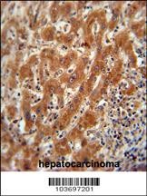
Pyruvate Kinase (PKM2) Antibody immunohistochemistry analysis in formalin fixed and paraffin embedded human hepatocarcinoma followed by peroxidase conjugation of the secondary antibody and DAB staining. This data demonstrates the use of the Pyruvate Kinase (PKM2) Antibody for immunohistochemistry.Application
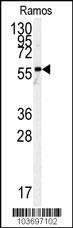
Western blot analysis of anti-h PKM2-N491 Pab in Ramos cell line lysates (35ug/lane).Application
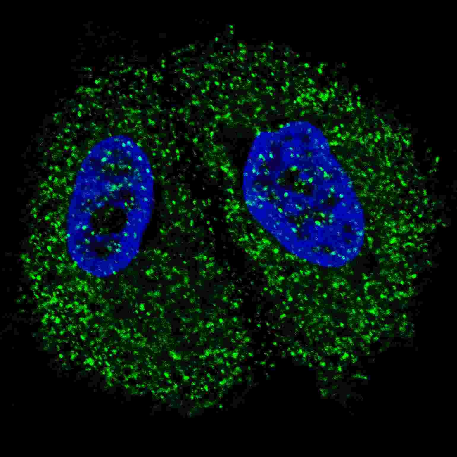
Fluorescent confocal image of MCF7 cells stained with Pyruvate Kinase (PKM2) antibody. MCF7 cells were fixed with 4% PFA (20 min), permeabilized with Triton X-100 (0.2%, 30 min). Cells were then incubated with Pyruvate Kinase (PKM2) primary antibody (1:200, 2 h at room temperature). For secondary antibody, Alexa Fluor 488 conjugated donkey anti-rabbit antibody (green) was used (1:1000, 1h). NucleiApplication
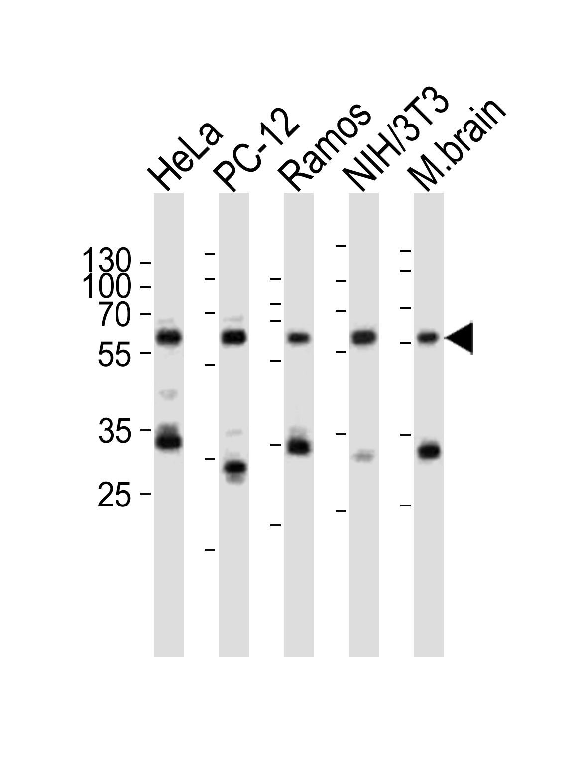
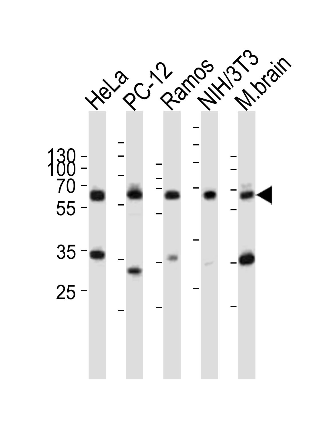
Western blot analysis of lysates from HeLa, rat PC-12, Ramos, mouse NIH/3T3 cell line, mouse brain tissue lysate(from left to right), using PKM2-N491 at 1:1000 at each lane.Application
Western blot analysis of lysates from HeLa, rat PC-12, Ramos, mouse NIH/3T3 cell line, mouse brain tissue lysate(from left to right), using PKM2-N491 at 1:1000 at each lane.| Product Name | Pyruvate Kinase Antibody |
|---|---|
| Antibody Type | Primary Antibodies |
| Product description | There are 4 isozymes of pyruvate kinase in mammals: L, R, M1 and M2. PKM2 is a pyruvate kinase that catalyzes the production of phosphoenolpyruvate from pyruvate and ATP. This protein has been shown to interact with thyroid hormone, and thus may mediate cellular metabolic effects induced by thyroid hormones. This protein has been found to bind Opa protein, a bacterial outer membrane protein involved in gonococcal adherence to and invasion of human cells, suggesting a role of this protein in bacterial pathogenesis.1) References for protein: |
| Immunogen | This Pyruvate Kinase (PKM2) antibody is generated from rabbits immunized with a KLH conjugated synthetic peptide between 476-505 amino acids from the C-terminal region of human Pyruvate Kinase (PKM2). |
| Clonality | Polyclonal |
|---|---|
| Isotype | Ig |
| Host Species | Rabbit |
| Tested Applications | IFIHC-PWB |
| For WB starting dilution is: 1:1000 For IF starting dilution is: 1:200 For IHC-P starting dilution is: 1:50~1:100 | |
| Species Reactivity | HumanMonkeyMouseRat |
| Concentration | 1mg/ml |
| Purification | Unpurified |
| Gene Symbol | PKM |
|---|---|
| Alternative Names | Pyruvate kinase PKM Cytosolic thyroid hormone-binding protein CTHBP Opa-interacting protein 3 OIP-3 Pyruvate kinase 2/3 Pyruvate kinase muscle isozyme Thyroid hormone-binding protein 1 THBP1 Tumor M2-PK p58 PKM OIP3 PK2 PK3 PKM2 |
| Molecular Weight(MW) | 58 kDa |
Application
Pyruvate Kinase (PKM2) Antibody immunohistochemistry analysis in formalin fixed and paraffin embedded human hepatocarcinoma followed by peroxidase conjugation of the secondary antibody and DAB staining. This data demonstrates the use of the Pyruvate Kinase (PKM2) Antibody for immunohistochemistry.
Application
Western blot analysis of anti-h PKM2-N491 Pab in Ramos cell line lysates (35ug/lane).
Application
Fluorescent confocal image of MCF7 cells stained with Pyruvate Kinase (PKM2) antibody. MCF7 cells were fixed with 4% PFA (20 min), permeabilized with Triton X-100 (0.2%, 30 min). Cells were then incubated with Pyruvate Kinase (PKM2) primary antibody (1:200, 2 h at room temperature). For secondary antibody, Alexa Fluor 488 conjugated donkey anti-rabbit antibody (green) was used (1:1000, 1h). Nuclei
Application
Western blot analysis of lysates from HeLa, rat PC-12, Ramos, mouse NIH/3T3 cell line, mouse brain tissue lysate(from left to right), using PKM2-N491 at 1:1000 at each lane.
Application
Western blot analysis of lysates from HeLa, rat PC-12, Ramos, mouse NIH/3T3 cell line, mouse brain tissue lysate(from left to right), using PKM2-N491 at 1:1000 at each lane.| Application Notes | For WB starting dilution is: 1:1000 For IF starting dilution is: 1:200 For IHC-P starting dilution is: 1:50~1:100 |
|---|
| Form | Liquid |
|---|---|
| Storage Instructions | Store at 4˚C for three months and -20˚C, stable for up to one year. As with all antibodies care should be taken to avoid repeated freeze thaw cycles. Antibodies should not be exposed to prolonged high temperatures. |
| Storage Buffer | Supplied in PBS with 0.09% (W/V) sodium azide. |


 Data sheet for OM289336